


 15 / 24

SUMMER 2015 ISSUE
RECURRENT UVE I T I S
Medical feature: equine
recurrent uveitis
What is equine recurrent uveitis?
Equine recurrent uveitis (ERU) is a very painful
condition that can lead to a permanent loss
of sight and, if left untreated, removal of the
eye (enucleation) is necessary. ERU is a
complex, immune-mediated disease, which
triggers inflammation in the uveal tract of the
eye (middle layer).
What causes Equine Recurrent Uveitis?
ERU usually begins with a bout of acute
uveitis. The patient is said to have ‘recurrent’
uveitis when they have had more than one
episode.
Triggers that set off ERU are: trauma (both
penetrating and blunt) or systemic disease.
ERU is a worldwide problem and specific
pathogens that have been attributed include
bacteria, in particular Leptospira species,
parasites (e.g. gastrointestinal worms) and
viral diseases such as equine herpes virus.
Clinical signs of acute anterior uveitis
●
A closed or partially closed eye
●
Excessive tears and sometimes a
mucopurulent discharge
●
An inflamed conjunctiva (conjunctivitis)
●
A cloudy eye due to a phenomenon
known as corneal oedema
●
In severe cases the eye may have a
creamy appearance with a red layer or
flecks in it. This is due to hypopyon and
hyphaema (pus and blood in the
front/anterior chamber of the eye).
●
A constricted pupil known as miosis
Clinical signs of chronic or recurrent
uveitis
●
A darkening of the iris, it can almost
appear black. A normally healthy brown
iris is bright in colour and, when observed
closely, is made up of different shades of
brown having a striped appearance.
●
Synechiae – this is where the iris, when
constricted (miosis) has stuck to the lens
capsule and, when it eventually relaxes
and dilates, bits of the iris remain
attached to the lens. This can leave black
flecks on the lens or even holes in the iris.
●
Other complications can occur such as;
cataracts, glaucoma and retinal
detachment
Diagnosis of ERU
●
Diagnosis is based on clinical signs and
history
●
A full clinical examination to look for
systemic disease that might be the cause
of ERU
Treatment of uveitis
Think of uveitis as a fire in the eye. It must
be put out immediately. ‘Fire extinguisher’
treatments are:
●
Topical anti-inflammatories – eye drops
(e.g. topical steroids)
●
Systemic anti-inflammatories – oral
medication
●
Mydriatics (eye drops to dilate the pupil)
●
Sub-conjunctival injections of steroid
may be given by your vet but extra
care must be taken to monitor that no
corneal ulcers are present or develop
similar to topical steroids
When treating uveitis, steps must be taken
to protect the affected eye from sunlight.
Medication to dilate the pupil will prevent
the pupil constricting in bright sunlight.
Keep the horse stabled during daylight hours
or sew a dark patch of cloth into a fly-mask
to protect against the sun.
Treatment and prevention of ERU
Initial treatment is the same as acute uveitis.
The treatment should persist for up to a
month after the initial signs were detected.
However, due to the propensity for ERU to
‘reignite’ without warning, it can become
increasingly difficult to manage the disease
so a ‘fire retardant’ approach is sometimes
required. Cyclosporin is a powerful
immunosuppressant. Small implants can be
placed in the eye under general anaesthetic;
slowly releasing the medication for
approximately three years. Patients treated
with a cyclosporin implant have a statistically
higher chance of retaining their eyesight.
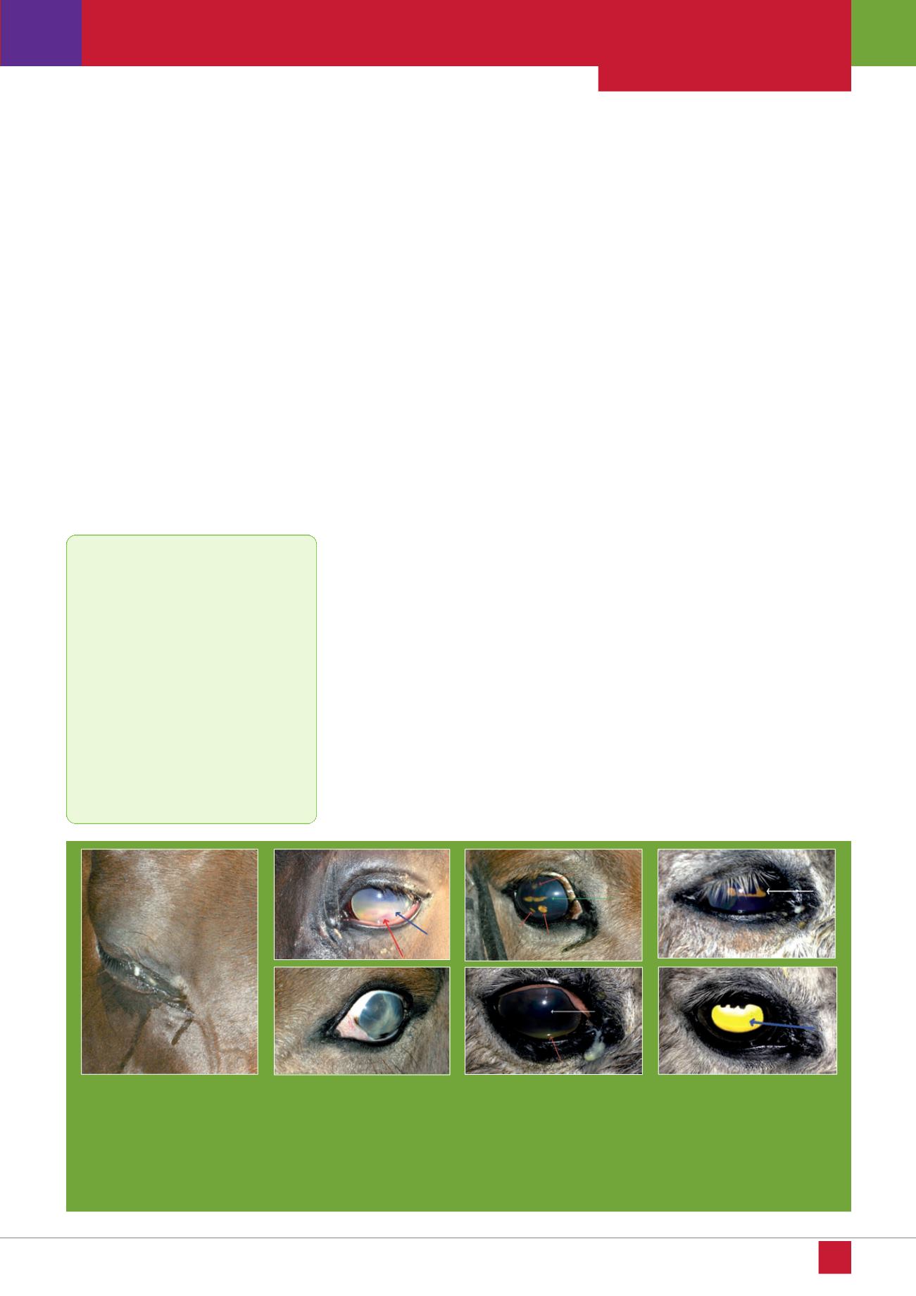
Figure 1
Figure 5
Figure 6
Figure 7
Key fact:
Whilst infections are commonly
implicated as a trigger of ERU, it is
the development of autoimmune activity,
the body’s defence mechanism against
these infections, which is likely to be
a major component of the process. In
laymen’s terms, ERU is an ocular
condition similar to recurrent airway
obstruction (RAO) in the horse or hay
fever and asthma in humans. These
are conditions where the body
overreacts to stimuli and the immune
response can cause more harm than
the original problem(s).
Figure 2
Figure 3
Figure 4
EQUINE MATTERS
14
Figure 1: a closed, painful eye with
tear staining
Figure 2: acute uveitis showing pus
(blue arrow) and flecks of blood
(red arrow) in the anterior chamber
of the eye
Figure 3: a shrunken, scarred blind
eye due to ERU
Figure 4: an eye suffering ERU. Note the dark iris and constricted pupil (green arrow) with small holes in the iris where
it stuck to the lens beneath (red arrows)
Figure 5: an eye with acute uveitis. Note the constricted pupil (white arrow) and small amount of sediment (pus – red
arrow) at the bottom of the eye
Figure 6: the eye one day after treatment. The pupil is now beginning to dilate (white arrow)
Figure 7: the same eye three days into treatment. The pupil is fully dilated and the yellow coloured vitreous, seen through
the pupil (blue arrow) can be a feature of animals developing uveitis due to an immune response to parasites. At the time
this horse was being treated for cyathostominosis (redworm).