


 12 / 24

11
EQUINE MATTERS
EYE D I SEASES
Veterinary surgeon
Aoife Byrne
XLEquine practice
Chapelfield Veterinary
Partnership Ltd
Equine
eye
diseases
The safety of a horse and its ability to do its work depends heavily on its
vision.
Whilst many horses can cope fairly well with compromised
vision, especially where this develops slowly, visual compromise
will not necessarily preclude the animal from being ridden.
Aoife Byrne DrMedVet MRCVS,
Chapelfield Veterinary Partnership Ltd
In this article five groups of
equine eye diseases will be
described.
These are:
1. Keratitis
2. Uveitis
3. Lens luxation
4. Cataracts
5. Retinal disease and
dislocation/detachment.
In spite of this, there are many horses that
work well even though they have obvious,
compromising eye disease.
The outward evidence of ophthalmic disease
is obvious when blepharospasm (excessive
blinking), epiphora (overflow of tears), eye
rubbing, head tilt, obvious asymmetry of shape
or size when compared to the normal eye,
changes in the clarity of the cornea and
obviously abnormal discharges are seen.
More subtle changes associated with
ophthalmic pain include downturned
eyelashes, drooping of the upper eyelid,
enophthalmos (eye drawn back into orbit)
and photophobia (sensitivity to bright light).
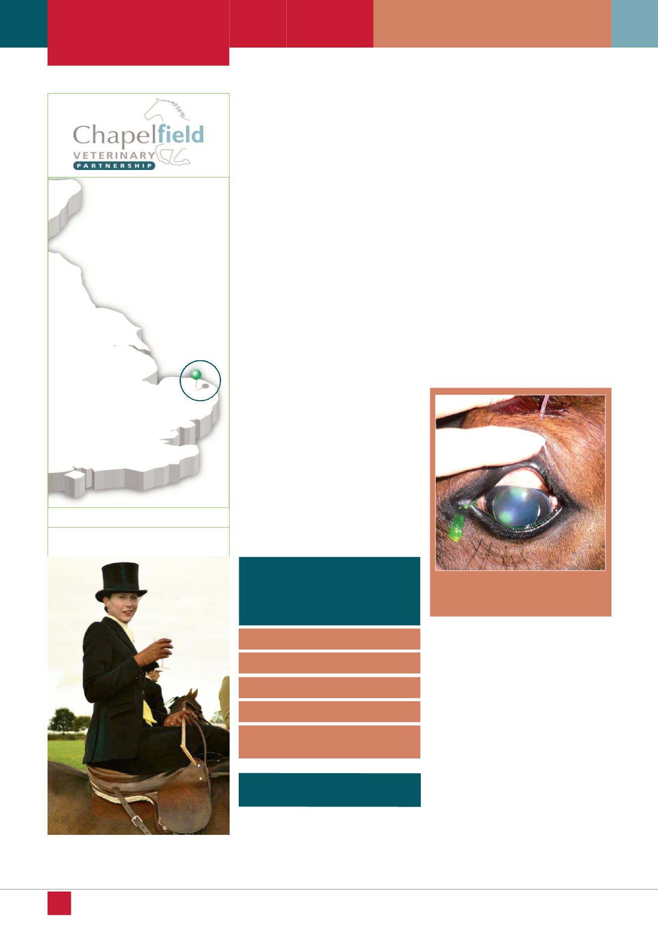
Corneal ulceration
(Figure 1)
is a potentially
sight threatening disorder requiring early
diagnosis, laboratory confirmation of
micro-organisms and appropriate therapy.
Viral, bacterial and fungal species may
be involved either as a primary cause
or as secondary infection and each requires
prompt therapy if serious ocular
complications are to be avoided.
Ulceration should be considered in every
acute or chronically painful eye and infection
should be considered in every corneal ulcer.
Fungal involvement s rare in the UK but
should be suspected with a history of corneal
injury with plant material or if the ulcer has
received prolonged antibiotics and has
shown no improvement.
Many early cases of ulcerative keratitis
present as minor corneal epithelial ulcers
or infiltrates with pain, blepharospasm,
epiphora and photophobia.
Keratitis
Figure 1: Superficial corneal ulceration
stained with fluoroscein dye