


 13 / 24

EQUINE MATTERS
12
SUMMER 2015 ISSUE
EYE D I SEASES
Cataracts & other lens conditions
The lens forms part of the focusing system
that delivers sharp images onto the retina
and has three zones which, from the centre,
are the nucleus, the cortex and the lens
capsule. A cataract is defined as any
opacity (cloudiness) within any of these
three layers. The position of opacities and
their size/extent will determine the amount
of visual impairment. Most horses appear
to cope well with ‘minor’ lens changes
however behaviour and athletic ability
are known to be affected by ‘significant’
cataracts.
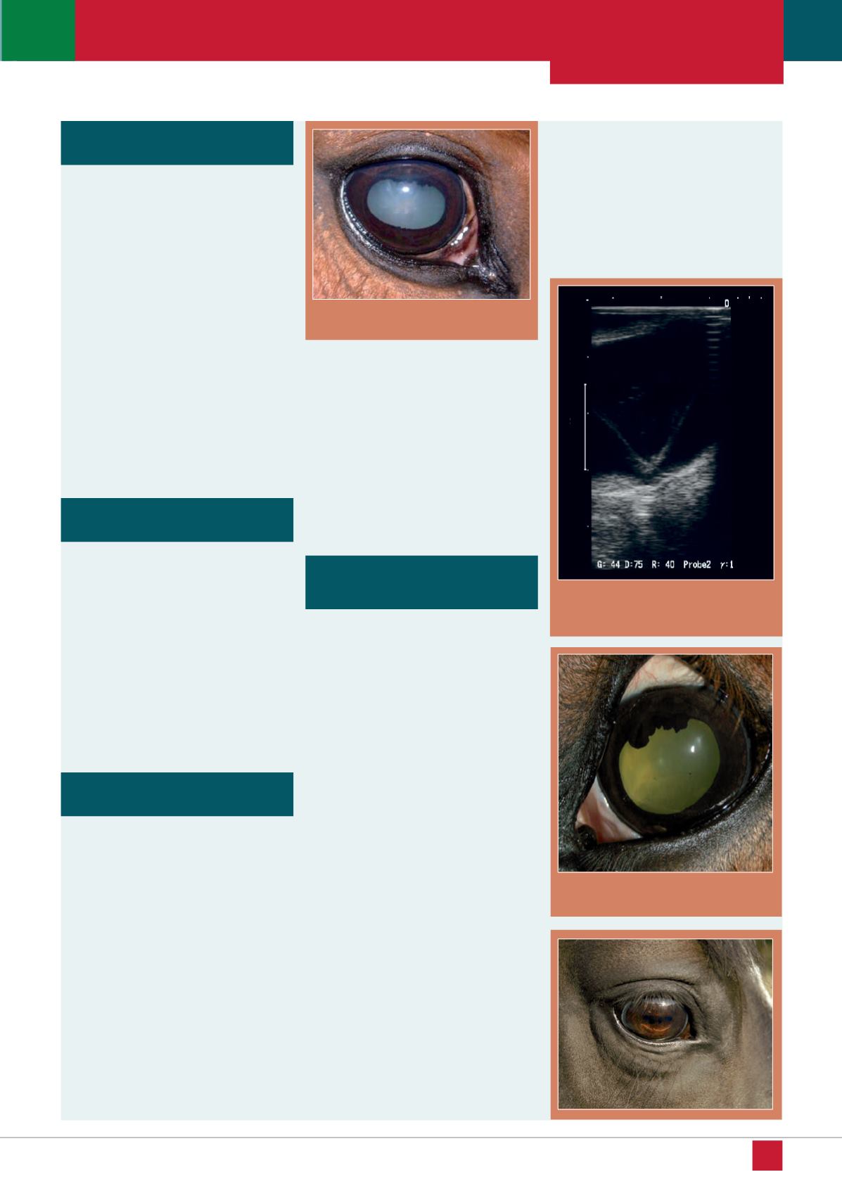
Cataracts
(Figure 2)
are categorised by
their level of maturity. Incipient/early
cataracts involve small areas of the lens
and do not affect vision. Immature cataracts
involve more of the lens with increasing
effects on vision. Mature cataracts involve
the entire lens and cause blindness.
Uveitis is inflammation of the middle
layer of the eye, the uvea. Uveitis can
be grouped into traumatic, reflex or
recurrent/persistent types. It can occur
as an intraocular primary event or as a
result of any other ocular disorder
(secondary/reflex uveitis). Immune
mediated equine recurrent uveitis (ERU)
is the most commonly recognised disease
entity of the equine eye. Uveitis is a
painful eye condition.
A range of clinical presentations may be
seen but in general the clinical signs are
non-specific inflammation of the uvea.
Treatment can be lengthy and complicated.
Prognosis is good with prompt diagnosis
and treatment of simple cases but
complications that interfere with vision
are common with delayed treatment and
severe cases.
Figure 2: Equine cataract
Due to a congenital defect in foals or
severe trauma in adults, the lens can luxate
forward or backwards from its normal
position. Movement of the iris from lens
contact, shallow or deep anterior
chambers, and aphakic (no lens) crescents
(edge of lens seen) might be present.
Cataract formation might also be noticed.
Dislocation of the lens into the vitreous
humour (gel between lens and retina) might
not necessitate surgery; however, movement
into the anterior chamber usually requires
removal to prevent secondary glaucoma
(increased intraocular pressure).
Uveitis
Cataracts block the visual image as they
increase in size, but don’t block light.
Congenital (present at birth) cataracts are
seen in foals, often in both eyes. In adult
horses, cataracts might be caused by trauma,
nutritional deficiencies or toxicities, or be
secondary to other conditions such as ERU.
An examination will determine if ERU is
also present; this is especially important when
cataract surgery is being considered, since
there is an increased risk of complications
and a poorer prognosis for vision when
uveitis is the cause of the cataract.
Lens luxation (dislocation)
Retinal disease and dislocation/
detachment
Chorioretinitis is inflammation of the
choroid and retina. It can be caused by
infectious agents, a poorly controlled
immune system, trauma or vascular
disease. It can be found with or without
ERU. It can be seen as focal "bullet-hole"
lesions, diffuse (spread out) lesions,
horizontal bands in the non-tapetum
(non-reflective back of eye) and
chorioretinal degeneration near the optic
nerve. Active chorioretinitis appears as
focal white spots with indistinct edges,
and as large, diffuse gelatinous grey
regions of retinal oedema (fluid swelling).
Inactive chorioretinitis can appear as
circular depigmented white regions with
hyper-pigmented (darkened) centres, or
large areas of depigmentation that appear
similar to the wings of a butterfly.
Congenital stationary night blindness
(CSNB) is found mainly in the Appaloosa,
and is inherited as a recessive trait.
Cases are also noted in Thoroughbreds,
Paso Finos, and Standardbreds. CSNB
appears to be caused by a failure of
neurotransmission in the middle retina.
Clinical signs include visual impairment
in the dark with (generally) normal vision
in daylight. There is behavioural
uneasiness and unpredictability at night.
Retinal detachment
(Figure 3)
is
separation of the layers of the retina,
which can be partial or complete. It is
associated with slowly progressive or
acute blindness. It can be congenital in
foals or acquired in adults and can
occur in one or both eyes. It can be a
complication of ERU
(Figure 4)
and
associated with congenitally small
eyes in foals, head trauma, wounds that
cause the cornea to rupture, cataract
surgery or secondary to intraocular
tumours.
Figure 3: Ultrasound image showing
detachment of the retina
Figure 4: ERU and retinal detachment