


 6 / 24

5
EQUINE MATTERS
David Rutherford BVM&S CertES(Orth) DipECVS MRCVS
Fellowes Farm Equine Clinic Ltd
OSTEOCHONDR I T I S
Veterinary surgeon
David Rutherford
XLEquine practice
Fellowes Farm
Equine Clinic Ltd
Osteochondritis dissecans
in horses
OCD is caused by a combination of factors:
●
Genetics - OCD is at least partially
inherited.
●
Rapid growth and large body size
●
Nutrition - diets high in energy or with
a mineral imbalance (usually low copper)
●
Hormonal imbalances - insulin and
thyroid hormones
●
Trauma - possibly a specific injury or
just during 'normal' exercise
Osteochondritis dissecans (OCD) affects 5-25% of all horses, but it is most common in
Thoroughbreds and Warmbloods and rare in ponies. It occurs when the joint surface of
young growing horses does not form properly, causing the cartilage and bone underneath
it to be irregular and weak. This can lead to the development of cartilage and bone flaps,
which either remain partially attached to the bone or break off and float around the joint.
These loose fragments irritate the inside of the joint causing joint swelling and moderate
lameness. Age at the development of signs can vary but 18 months to 4 years is most
common – often joint swelling and lameness initially occurs when a young horse begins work
for the first time. OCD can occur in all joints, but is most common in the stifle and hock.
OCD might be suspected when a large
young horse develops lameness and a
swollen joint, and is easily confirmed with
x-rays. Often loose bone fragments can be
seen, or if just the cartilage is involved, we
just see a flattened area on the joint surface.
OCD is commonly present within the same
joint in the other leg even if there are no
signs, so x-rays of the other leg should
always be taken.
Occasionally, OCD might be treated by joint
medication alone, but the optimum treatment
is the removal of loose bone fragments and
diseased cartilage by keyhole surgery
(arthroscopy). A camera the size of a pencil
is inserted into the affected joint through a
1cm incision. The joint is distended with
sterile saline solution or carbon dioxide gas,
and the joint surface can be examined using
the image projected onto a television monitor.
The loose fragments and soft diseased
cartilage are located and removed using
grasping instruments placed through a second
small incision. Any soft bone under the
fragments can then be scraped away before
the joint is flushed clean and the skin sutured
closed. Arthroscopy is usually performed by
specialist equine surgeons under general
anaesthetic in an equine operating theatre,
but it is occasionally possible for surgery to
be undertaken in the standing sedated horse.
It is not a cheap undertaking with the cost of
investigations, surgery and immediate
aftercare being approximately £2500-3500.
After surgery horses will typically be stable
rested for about 3 weeks and then undertake
a steady return to exercise over the next few
months. Often the affected joint(s) will be
injected with cortisone and lubricants 4-6
weeks after surgery to remove any residual
inflammation. Alternatively biologic therapies
such as platelet rich plasma (PRP) or stem
cells can be injected to help stimulate healthy
cartilage to cover the exposed bone surface.
The prognosis for a full athletic career
following OCD surgery is good to excellent
in most cases with the best results achieved
with smaller injuries and when horses are
treated at a young age.
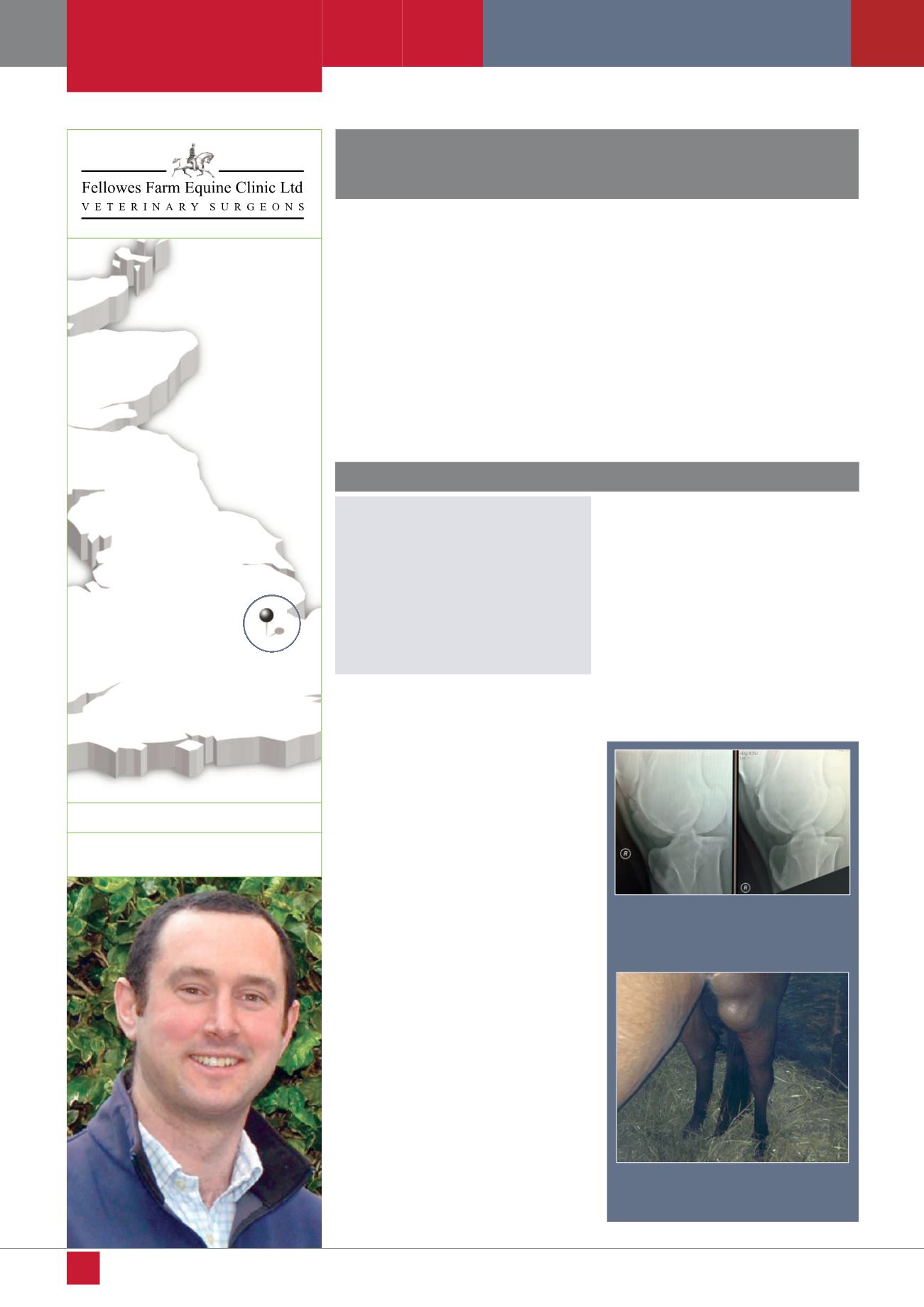
Stifle effusion associated with an
OCD lesion
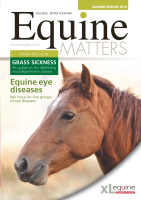
X-rays of a horse’s stifle show an OCD
fragment at the front of the joint before
(left) and after (right) surgery to remove it