


 15 / 20

repeating images. Depending on the reasons for taking
the radiographs, removal of shoes may or may not be
necessary. For example, performing radiographs with
the shoes in place can be useful when assessing foot
balance and conformation as they allow accurate
identification of the weight-bearing surface and the
position of the shoe in relation to the hoof capsule and
pedal bone. However, if a source of lameness has been
localised to the foot, removal of the shoes may be
required to obtain the additional views necessary to
fully evaluate the foot.
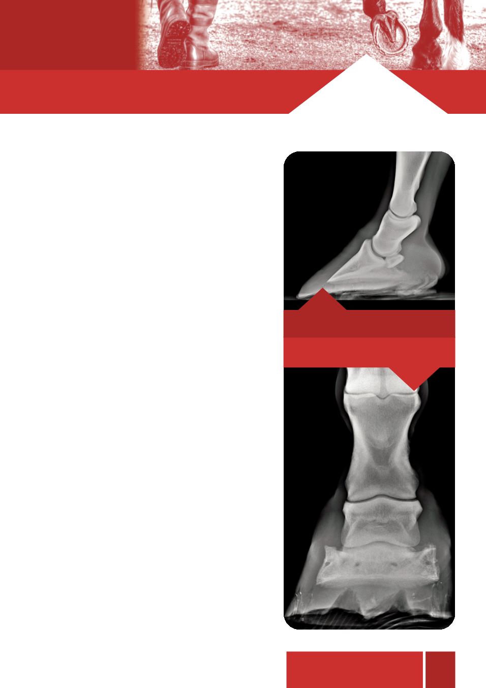
Two views are usually required to fully assess balance
of the feet, one lateral (taken from the side of the limb),
and one dorsopalmar (taken from the front). Bone
alignment, heel and toe length and sole depth can all
be assessed on the lateral radiograph with particular
attention being paid to the alignment of the pastern and
pedal bones and their relationship to the hoof capsule, the
so called ‘hoof pastern axis’. A broken-back hoof pastern
axis is a common abnormality, particularly in warmbloods,
causing increased loading of the heels, straining the deep
digital flexor tendon and supporting ligaments of the
navicular bone. The dorsopalmar view is used to assess
joint alignment, foot symmetry and the length of both the
medial (inner) and lateral (outer) hoof walls. Excessive
length on either side can affect the rest of the limb by
causing uneven loading of joints, causing stress on
collateral ligaments or leading to osteoarthritis.
Routine radiographic imaging of the foot can be an
invaluable aid to trimming, shoeing, injury prevention, and
treatment of injury, whether the goal is to simply keep the
horse healthy and happy at pasture, or to facilitate optimal
performance of a competitive equine athlete.
14
...Keep one step ahead
Broken back hoof pastern axis.
Mediolateral imbalance.