


 14 / 20

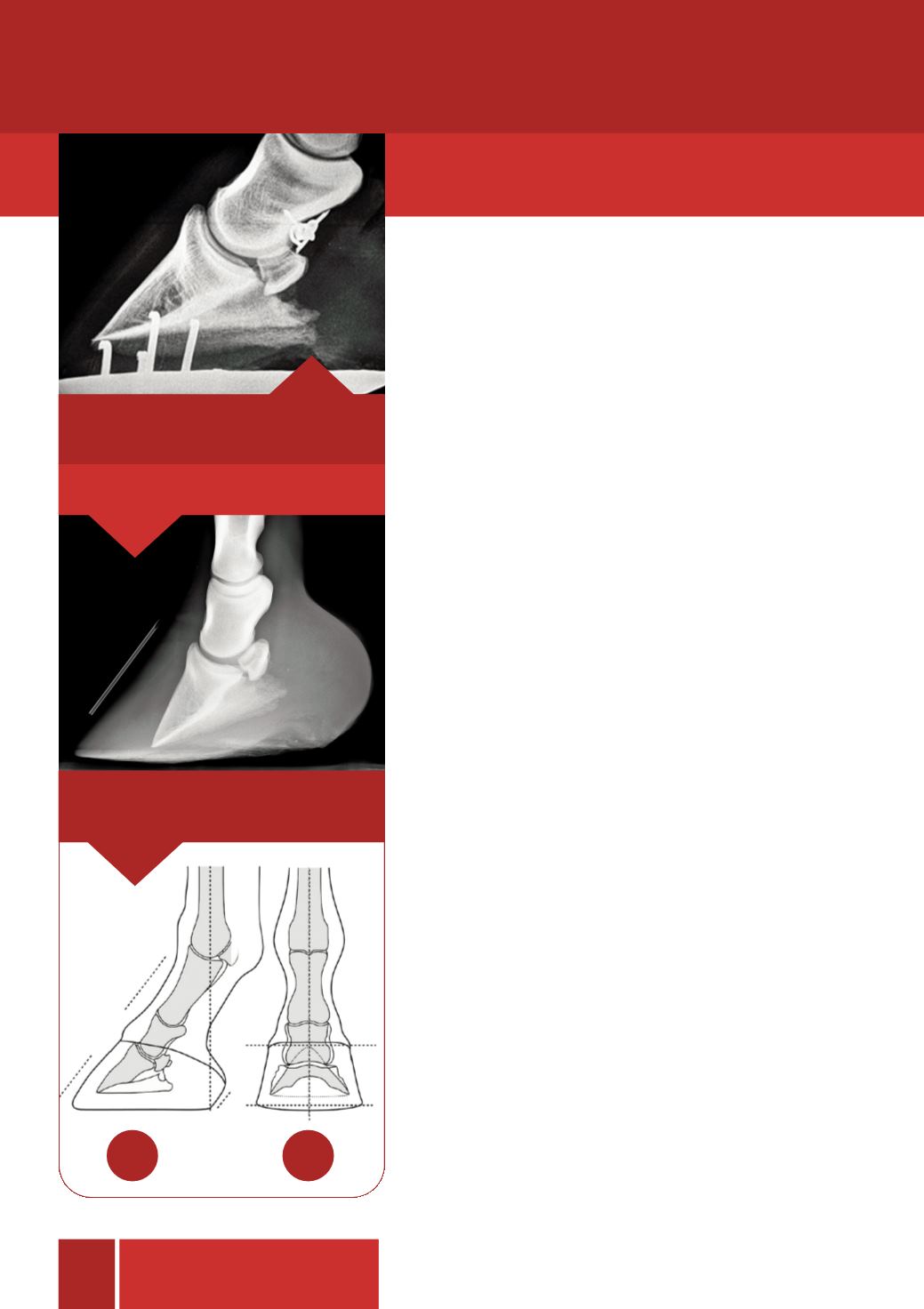
Foot pain is a major cause of equine lameness and
radiography is an important diagnostic tool in identifying
the location, extent and severity of any injury or disease
within the foot. It’s most important role is to give
information about bones and joints, however, it can
provide information about soft tissues such as tendons
and ligaments, particularly where they attach to bone.
Some common causes of foot pain which may be
identified with the aid of radiography include pedal
bone fractures, osteomyelitis (infection of the bone),
osteoarthritis, laminitis and navicular disease.
In the absence of lameness, radiographic evaluation of the
feet can also be used to provide information about foot
balance and conformation. An animal which exhibits poor
foot conformation, imbalance or abnormal patterns of
growth will be at significantly greater risk of developing
lameness as a result of abnormal forces on the bones and
soft tissues leading to injury. Consequently radiography
should be considered a powerful tool, not only in the
diagnosis, but also in the prevention of lameness by
providing information about the specific farriery
requirements of an individual horse. Using radiographs,
vet and farrier can work together as a team to maintain
or improve a horse's soundness and performance.
The widespread use of mobile digital x-ray machines
means that radiography of the feet is a straightforward
procedure but it can take time to prepare and position the
feet correctly so as to avoid the need, risk and expense of
Radiography
of
the equine foot
13
Foot care
The equine foot is one of, if not, the most common
area of interest for radiographic evaluation.
Equine
foot care
Pedal bone rotation.
Image A
- Hoof pastern axis.
Image B
- Mediolateral hoof balance.
Unusual barbed wire wound to the coronary
band.
A
B