


 5 / 20

04
...Keep one step ahead
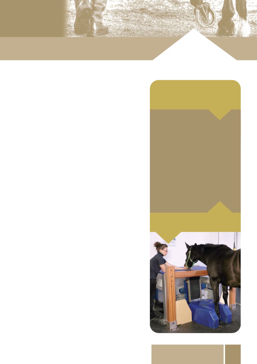
MRI scans
can be an important tool in
diagnosing the cause of a foot lameness.
Investigation of foot lameness
All investigations should begin with a good history where
information such as recent injuries, changes in farrier,
changes in feeding and exercise levels can all be
discussed. A visual inspection will evaluate the stance,
conformation and symmetry; which can give vital clues to
possible areas of trouble.
Palpation (feeling) of the limb and foot will be performed;
heat or an increase in digital pulses can help in identifying
foot inflammation. The pulses may be easiest to feel along
the back of the fetlock and a comparison should be made
with the opposite leg. The application of hoof testers,
which are designed to apply pressure in selected areas of
the foot such as the frog or sole, can help localise foot
pain and may assist in diagnosing a bruised sole, an
abscess, nail bind, laminitis or heel pain.
In cases where the source of lameness is unclear or
needs confirmation the next step is the localisation of the
pain using a nerve block or joint block. Local anaesthetic
is placed around a nerve or into a joint or bursa to numb
the area. Once an area is ‘blocked’; if the horse becomes
sound we then know that area is relevant to the cause of
the lameness. We then need to image the area to identify
any abnormalities. This is most commonly done using
x-rays. We can visualise fractures, navicular degeneration,
osteoarthritis in the coffin joint, pedal bone changes
such as pedal osteitis or changes in position of the pedal
bone such as in laminitis. Sometimes if the lameness is
caused by damage to soft tissue structures, very little may
be seen on the x-rays and other imaging techniques need
to be used. Other techniques used include ultrasound
imaging. This can be used as well as nuclear scintigraphy
(bone scan), magnetic resonance imaging (MRI scan),
computed tomography (CT scan), thermography and
contrast venography.
history
visual examination
palpation (hands and hoof testers)
nerve and joint blocks
radiography
contrast venography
ultrasound
nuclear scintigraphy (bone scan)
MRI
CT
thermography
Investigation of foot
lameness may involve: