


 4 / 20

Anatomically the foot is a complex structure with bones
(pedal, pastern and navicular) and numerous tendons
and ligaments that insert into these bones. The coffin
joint includes a joint capsule and joint surfaces that are
potential sites for inflammation. The navicular bursa is a
fluid cushion which protects the deep flexor tendon as it
runs over the navicular bone and can be involved in some
lameness cases. The laminae or interconnected ‘fingers’
are responsible for supporting a significant amount of the
horse’s weight and maintaining the pedal bone in the
correct position inside the hoof capsule. Cases of foot
lameness may involve more than one of these structures
at a single time. These structures are mostly hidden within
the hoof capsule so can be hard to visualise; therefore
associated disease or injury can be difficult to diagnose.
The foot as a cause
of
lameness
03
Foot care
Lameness can be described as a failure
in normal motion with a deviation from the
normal gait. Forelimb foot lameness is
more common than hindlimb lameness.
The centre of gravity of a horse is nearer
the front of the horse and at certain times
during the stride, huge forces are exerted
down the limbs, through the foot to the
ground. If we consider the small size of the
horse’s foot in relation to their body size, it
is not surprising that foot lameness is
very common.
Equine
foot care
The anatomy of the foot
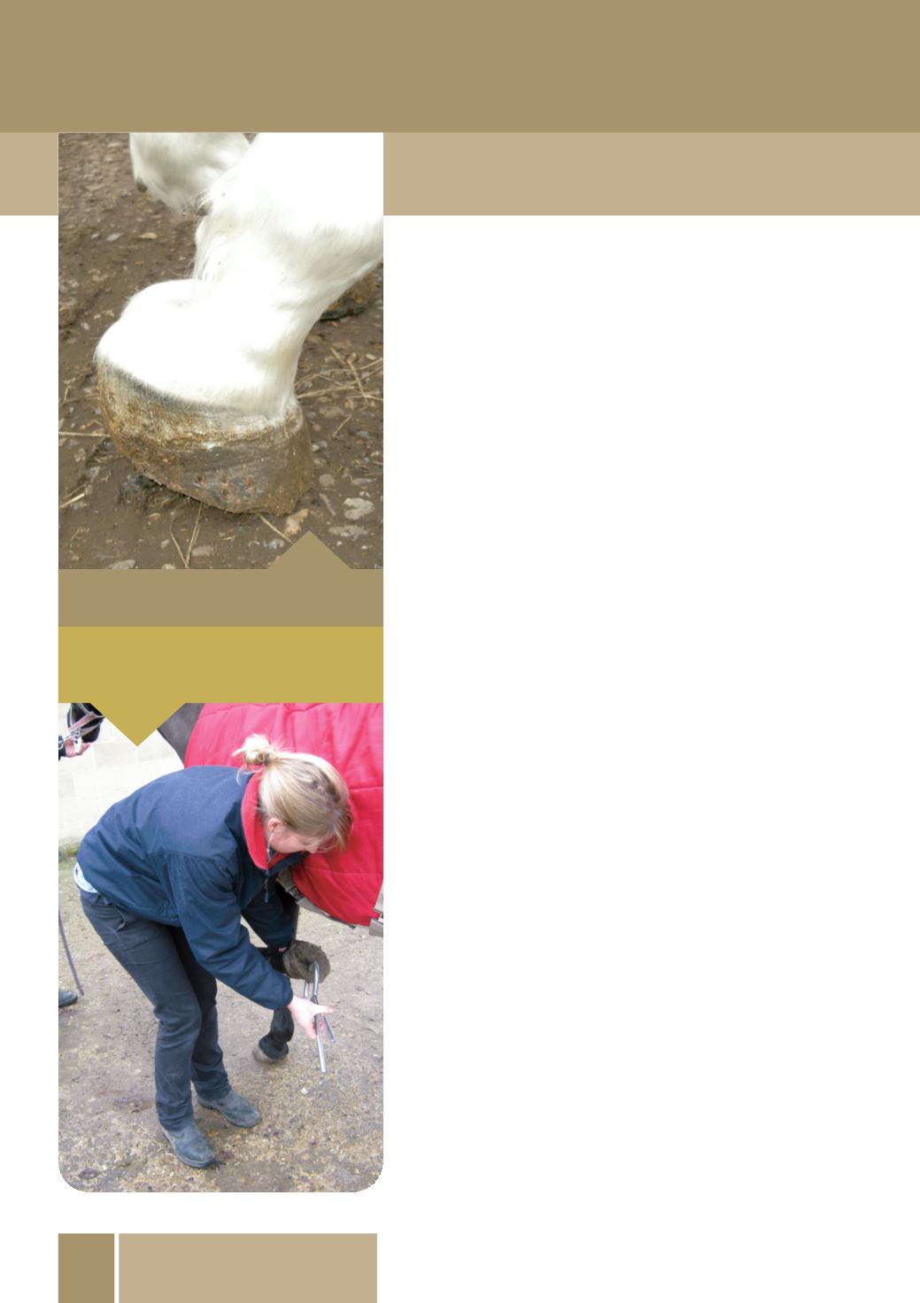
Hoof testers being applied to a horse’s foot
for lameness investigation.
Horse lifting heel off the ground due to pain.