


 21 / 24

SUMMER 2016 ISSUE
EQUINE MATTERS
20
Veterinary Surgeon
Andrew McDiarmid
XLEquine Practice
Clyde Veterinary
Group
ANDREW MCDIARMID, BVM&S CertES(Orth) MRCVS
CLYDE VETERINARY GROUP, LANARKSHIRE
ORTHOPAEDIC INJURY
Ruby was a well-developed, healthy foal and
lived what appeared a relatively normal life
for her first year. Unfortunately, in 2013
Cathy noticed that Ruby had started to
appear uneasy on her limbs, particularly her
hindlegs. She called her local vet to assess
the horse, who immediately recognised that
Ruby was showing signs of weakness and
ataxia (neurological weakness) whereby the
limbs were not co-ordinated. Ruby was
X-rayed and diagnosed with cervical
compressive myelopathy or ‘Wobblers
syndrome’. This is a developmental
abnormality where the bones of the neck
develop poorly and compress the spinal
cord; therefore the control of movement is
severely compromised and the horse becomes
wobbly. Due to a very poor prognosis in the
vast majority of cases, most owners elect to
have their animal euthanased. The vet knew
how important Ruby was to Cathy and how
her birth had helped her recover from her
own illness, so he brought to Cathy’s
attention that occasionally some horses may
be appropriate candidates for surgery.
Cathy was very keen to try this option, and
admitted the filly to Clyde Veterinary Group
Equine Hospital for further investigations to
determine if surgery was an option.
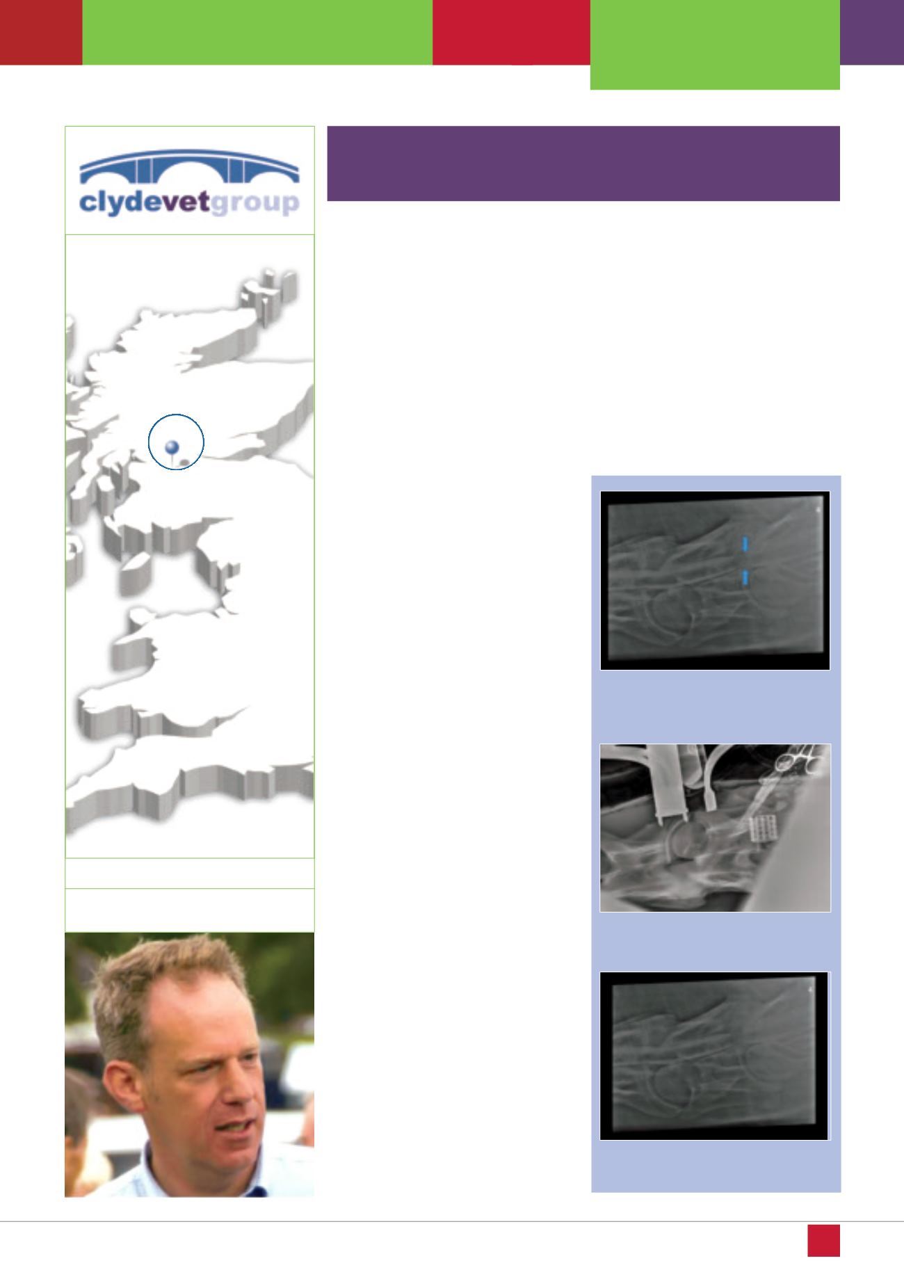
Ruby underwent a myelogram whilst under
general anaesthetic which recognised that
there was compression at two sites in the
neck between the fifth (C5) and the sixth
(C6) and the sixth and seventh (C7) cervical
vertebrae
(figure 1).
Andrew McDiarmid
consulted with members of the Liphook
Equine Hospital and the decision was
made to undertake the very risky neck
fusion surgery.
The three hour operation to insert two
stainless steel Bagsey baskets between the
two affected sites C5-C6 and C6-C7 was
undertaken without major complications
(figure 2)
and more critically, Ruby stood up
in the padded recovery room without issue.
Ruby returned home for a prolonged period
of rehabilitation over the next six months
(figure 3)
and now three years later, she
lives a relatively normal life with the ataxia
barely detectable. Indeed, to the untrained
eye she could be observed as a normal
horse grazing in the field.
Thankfully, Cathy’s health has also fully
returned and she now has the pleasure of
having Ruby at home behaving like any other
horse of her age.
Cathy had always longed to breed a foal and due to her unfortunate ill health
this dream became even more important to her. She was overwhelmed
therefore, with the birth of a fit, well bred filly foal ‘Ruby’ in 2012.
Figure 1. Spinal cord compression seen
using dye around the spinal cord
(myelogram)
Figure 2. Intraoperative X-ray of the second
basket being placed in the neck
Figure 3. Ruby's neck X-ray 6 months after
surgery
HAPPY END INGS