


 15 / 24

SPRING 2016 ISSUE
EQUINE MATTERS
14
D I ARRHOEA
Acute watery diarrhoea in a foal or adult
horse should not be ignored and warrants
discussion with your vet. One of the main
functions of the horse’s colon is to reabsorb
water, therefore profound diarrhoea usually
suggests significant disease of the colon.
This is often called ‘colitis’, especially if
inflammation of the colon wall has occurred.
Dehydration can rapidly occur due to the
ensuing massive water and electrolyte losses,
possibly leading to endotoxaemia or ‘septic
shock’
(figure 3)
. Horses can lose up to
50-100 litres of fluid in 24 hours due to
diarrhoea and so can deteriorate very quickly.
Following initial digestion in the stomach and
small intestine, food material passes into the
colon and caecum - a huge voluminous
reserve containing millions of ‘friendly’
bacteria. It is here where fermentation and
digestion of plant material occurs in the horse
that otherwise would be indigestible and a
wasted food source. It is also at this location
that fluid is secreted and absorbed to
maintain intestinal water regulation of the
horse. Therefore, when there is damage to
the colon wall and it can no longer regulate
this balance, diarrhoea occurs.
There are multiple causes of diarrhoea in
the adult horse and often, despite repeated
clinical examinations and extensive
diagnostics, a definitive diagnosis can be
frustratingly difficult to identify. However,
treatment is often similar and therefore can
be initiated before a specific diagnosis is
made. Causes of diarrhoea in the adult horse
include infective agents, such as
Salmonella
spp. or Clostridial bacteria and parasitic
infections, most commonly small strongyles
(redworm/cyathostomins). Non-infectious
causes include recent antibiotics or
non-steroidal anti-inflammatory administration,
toxin or sand ingestion. Sometimes anxiety
alone in a nervous or fractious animal will
lead to the temporary production of
watery faeces.
Diagnostic investigations include assessing
faecal material for larval stages of small
strongyles (redworm), bacterial culture and
DNA analysis of faecal samples.
Ultrasonography of the intestinal wall can be
very useful for assessing and measuring colon
wall inflammation and thickening, and may
help specify a diagnosis. For example, in
cases of diarrhoea related to recent
nonsteroidal anti-inflammatory drug (NSAID)
administration (e.g. ‘bute), a specific
syndrome of colitis of the right upper colon
occurs (known as right dorsal colitis), which
can be identified ultrasonographically. Bloods
are often taken to assess the degree of
inflammation present, to help rule in or out
infectious disease, and to assess the degree
of dehydration.
If a causal agent is identified, treatment
can be tailored more specifically, however
general treatment principles include
correction of dehydration and restoration
of normal electrolyte balance. This can be
achieved through oral fluid supplementation
or, if required, via an intravenous drip.
The inflammation may be severe enough for
protein to be lost across the intestinal wall
resulting in low protein levels within the
blood. Clinically this may be apparent as
oedema or fluid swelling of the lower legs
and under the belly
(figure 4)
. In very
severe cases, intravenous plasma may be
needed to restore blood protein levels.
Use of antibiotics in diarrhoea is controversial
and generally not advised as their use can
directly result in diarrhoea. Anti-inflammatories
will often be administered for both treating
the pain associated with acute diarrhoea
and also for controlling the inflammation;
however, they must be used carefully, as they
can further compromise the bowel health and
lead to other further complications in a
dehydrated patient.
For mild cases which remain bright and
eating without systemic signs, a course of
probiotics alone may be utilised to help
restore hindgut bacterial health, although
there is little scientific evidence to support
their use. Other additional treatments may
include bio-sponge, an oral product which
can help support a healthy intestinal function
and can absorb toxins from the bowel.
The prognosis for a mild case of diarrhoea is
generally good, although more severe cases
of colitis carry a far more guarded prognosis.
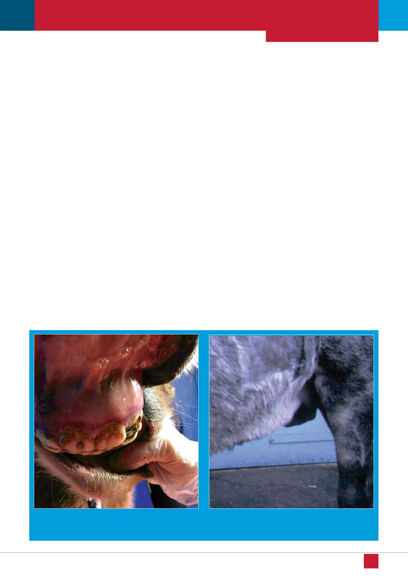
Figure 3.
Gums that are deep red with a purple ring are a hallmark
feature of septic shock
Figure 4.
Oedema (tissue fluid) accumulation under the belly in a
horse with low blood protein due to redworm (small strongyle)
associated diarrhoea