Basic HTML Version


LAMENESS
Lameness
can be described as a failure in normal motion with a deviation
from the normal gait. Forelimb foot lameness is more common than hindlimb
lameness. The centre of gravity of a horse is nearer the front of the horse and at
certain times during the stride, huge forces are exerted down the limbs, through
the foot to the ground. If we consider the small size of the horse’s foot in relation
to their body size, it is not surprising that foot lameness is very common.
The
foot
as a cause
of lameness
Graham Hunter BVM&S GPCert(EqP) CertEP CertAVP(ESO) MRCVS,
Ardene House Veterinary Practice
The anatomy of the foot
Anatomically the foot is a complex structure
with bones (pedal, pastern and navicular)
and numerous tendons and ligaments that
insert into these bones. The coffin joint
includes a joint capsule and joint surfaces
that are potential sites for inflammation. The
navicular bursa is a fluid cushion which
protects the deep flexor tendon as it runs
over the navicular bone and can be involved
in some lameness cases. The laminae or
interconnected ‘fingers’ are responsible for
supporting a significant amount of the horse’s
weight and maintaining the pedal bone in
the correct position inside the hoof capsule.
Cases of foot lameness may involve more
than one of these structures at a single time.
These structures are mostly hidden within the
hoof capsule so can be hard to visualise;
therefore associated disease or injury can
be difficult to diagnose.
All investigations should begin with a good
history where information such as recent
injuries, changes in farrier, changes in
feeding and exercise levels etc. can all be
discussed. A visual inspection will evaluate
the stance, conformation and symmetry;
which can give vital clues to possible areas
of trouble.
Palpation (feeling) of the limb and foot
will be performed; heat or an increase in
digital pulses can help in identifying foot
inflammation. The pulses may be easiest to
feel along the back of the fetlock and a
comparison should be made with the
opposite leg. The application of hoof testers,
which are designed to apply pressure in
selected areas of the foot such as the frog
or sole, can help localise foot pain and
may assist in diagnosing a bruised sole, an
abscess, nail bind, laminitis or heel pain.
In cases where the source of lameness is
unclear or needs confirmation the next step
is the localisation of the pain using a nerve
block or joint block. Local anaesthetic is
placed around a nerve or into a joint or
bursa to numb the area. Once an area is
‘blocked’; if the horse becomes sound we
then know that area is relevant to the cause
of the lameness. We then need to image the
area to identify any abnormalities. This is
most commonly done using x-rays. We can
visualise fractures, navicular degeneration,
osteoarthritis in the coffin joint, pedal bone
changes such as pedal osteitis or changes
in position of the pedal bone such as in
laminitis. Sometimes if the lameness is
caused by damage to soft tissue structures,
very little may be seen on the x-rays and
other imaging techniques need to be used.
Many other different techniques are used to
look at different tissues and structures in the
foot. Ultrasound imaging can be used as
well as nuclear scintigraphy (bone scan),
magnetic resonance imaging (MRI scan),
computed tomography (CT scan),
thermography and contrast venography.
Investigation of foot lameness
may involve:
l
history
l
visual examination
l
palpation (hands and hoof testers)
l
nerve and joint blocks
l
radiography
l
contrast venography
l
ultrasound
l
nuclear scintigraphy (bone scan)
l
MRI
l
CT
l
thermography
Investigation of foot lameness
9
EQUINE MATTERS
Veterinary Surgeon
Graham Hunter
XLVets Equine Practice
Ardene House Vet
Practice Ltd
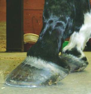
A long toe low heel conformation. This
horse is shod with poor support at the
back of the foot