Basic HTML Version


COL I C
Veterinary Surgeon
Julian Rishworth
XLVets Equine Practice
The Minster Veterinary
Practice
Julian Rishworth BVetMed MRCVS
, The Minster Veterinary Practice
3. Apply 2-3 layers of rolled cotton
wool to the limb, for padding and
further protection.
CASE REPORT...
Colic
requiring surgical
treatment
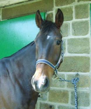
Oakley
is a 10 year old part
bred thoroughbred gelding, he
first showed colic symptoms one
evening, and his usual vets found
an impaction of the pelvic flexure
and treated it appropriately.
Unfortunately, Oakley continued to show
signs of colic the following day, despite the
treatment, which is not entirely unusual with
impactions but what was unusual was the
presence of distended loops of small intestine
on rectal examination. When further treatment
didn’t resolve the colic and Oakley continued
to display colic signs, the decision was made
to refer Oakley for further investigations.
On arrival at the clinic, Oakley had a
very mild elevation of heart rate and his
temperature was normal but his respiratory
rate was high at 40 breaths per minute. The
colour of his mucous membranes (gums) was
normal. There were still distended loops of
small intestine on rectal examination as well
as an impaction in the left colon. Scanning
the abdomen showed the distended small
intestine with more fluid surrounding the
intestines than would normally be expected.
A sample of this fluid was obtained from the
lower part of the abdomen and this showed
that the fluid was more red and cloudy than
normal; the normal fluid should be pale
yellow and clear. This was sufficient indication
that there was a problem in the abdomen that
needed surgery to correct but further blood
tests helped confirm this and in particular the
lactate levels showed that there was a
reasonable prognosis for a problem that
could be corrected by surgery.
Oakley was prepared for theatre immediately
by placing a catheter into the jugular vein in
the neck, and antibiotics and pain relief were
given before the start of surgery. Oakley was
anaesthetised and on the surgical table within
about one hour of his arrival at the clinic.
The surgical incision is made in the midline
base of the abdomen, just in front of the
umbilicus (belly button). Once in the
abdomen, the loops of distended small
intestine were obvious but the caecum was
very empty. The problem was identified as a
twist in the last part of the small intestine. This
had slowed the passage of food material and
partly compromised the blood supply to this
section of the bowel. This had resulted in the
small intestinal wall swelling, further stopping
food material completely, hence the empty
caecum and impacted material in the large
bowel, deprived of the important fluid which
would normally come from the small intestine.
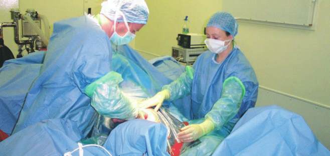
Surgery was performed to remove the
damaged section of small intestine and
create a new ‘join’ between the new end
of the small intestine and the caecum. In all,
Oakley had 16 feet of small intestine
removed in a surgery that took just under
three hours. After replacing the repaired
intestine back into the abdomen and repairing
the abdominal incision, Oakley made a good
recovery from anaesthesia. Post-operative
recovery was unremarkable. After seven
days in the clinic with antibiotics, pain relief,
intravenous fluid therapy and a careful
re-introduction to feed and water; Oakley
was discharged back to his owners for a
further three months box rest to allow the
surgical wound to fully heal. Oakley’s case is
a good example where prompt decisions by
his regular vets to recognise that this was
more than a regular pelvic flexure impaction
meant that surgery was more likely to be
successful with fewer complications.
Oakley
SUMMER 2013 ISSUE
EQUINE MATTERS
8
Oakley had 16 feet of small intestine removed at surgery