Basic HTML Version


LAMENESS
SUMMER 2013 ISSUE
EQUINE MATTERS
10
Treatment of foot lameness obviously relates
to the cause. In some cases specific
treatment may be indicated e.g. a foot
abscess needs to be drained and poulticed.
No matter what the diagnosis is, in foot
lameness cases and indeed many other
causes of lameness, foot trimming and
balancing are fundamental to successful
resolution of lameness.
Medical treatments used include joint or
bursal injections of anti-inflammatories or
compounds which assist with joint repair and
maintenance; these can be used in cases of
coffin joint or navicular bursal inflammation.
Tiludronic acid can be used in cases where
alteration in bone modelling is required, such
as in some navicular disease cases. Oral
anti-inflammatory pain relief such as ‘bute’
will also frequently form part of treatment
protocols in horses with foot lameness.
Surgery may be performed to obtain more
information as well as used in treatment.
Coffin joint arthroscopy and navicular
bursoscopy can be used to see inside these
synovial structures helping to visualise
damaged or affected tissue. During surgery
‘tidying up’ of damaged tissue can also be
performed. Other surgical procedures may
include cutting out of tissue, such as
keratomas (a type of benign tumour within
the foot) or infected areas of pedal bone.
As a last resort, neurectomies can be
performed i.e. cutting nerves to remove
pain sensation from the feet of chronically
lame horses.
Diagnosing the foot as the cause of the
lameness is relatively straight forward; yet
identifying affected structures and tissues with
reliable specificity remains the challenge.
Fortunately treatment strategies have
improved and diagnostic procedures exist
now which can help solve even the most
elusive of problems. Most foot lameness,
even when originating from the navicular
region; does not carry the poor prognosis
that was previously given in these cases.
Early diagnosis and perseverance in
treatment is frequently the key to success.
The treatment and management
of foot lameness
Examples of surgical treatments
used in foot lameness:
Examples of non- surgical
treatments used in foot
lameness:
l
palmar digital neurectomy;
l
desmotomy of the inferior check ligament;
l
arthroscopy of the coffin joint;
l
bursoscopy of the navicular bursa.
l
desmotomy of the collateral ligaments
of the navicular bone;
l
foot trimming;
l
remedial farriery;
l
NSAIDs (e.g. ‘bute’);
l
tiludronate;
l
isoxuprine;
l
intramuscular joint therapy;
l
extracorporeal shock wave therapy.
l
injection of corticosteroids, hyaluronic
acid, or regenerative therapies into the
coffin joint or navicular bursa;
A few examples of causes
of foot lameness:
l
nail bind/prick;
l
foot imbalance;
l
foot abscess;
l
thrush;
l
solar bruising;
l
corns;
l
sheared heals;
l
canker;
l
puncture wounds;
l
coronary band and hoof wall lacerations;
l
hoof wall separation (white line disease);
l
pedal bone infection;
l
keratoma;
l
navicular disease;
l
coffin joint osteoarthritis;
l
laminitis;
l
deep digital flexor tendonitis;
l
coffin joint collateral ligament injury.
l
quittor;
l
pedal bone fractures;
l
navicular bone fractures
l
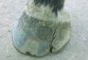
hoof wall cracks
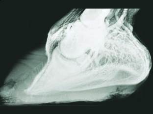
Contrast venogram of a chronic laminitic
foot showing good circulation
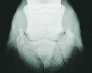
X-Ray showing a pedal bone fracture