


 6 / 24

5
EQUINE MATTERS
ANTIMICROBIAL
RESISTANCE
Veterinary surgeon
Dr Imogen Burrows
XLEquine practice
Cliffe Veterinary
Group
A complicated castration
Dr Imogen Burrows BVetMed CertAVP(EM) MRCVS,
RCVS Advanced Veterinary Practitioner in Equine Medicine
Cliffe Equine Clinic
Castration is one of the commonest surgical procedures performed in
veterinary practice and is generally straightforward, however as with
any surgical technique complications can and occasionally do occur.
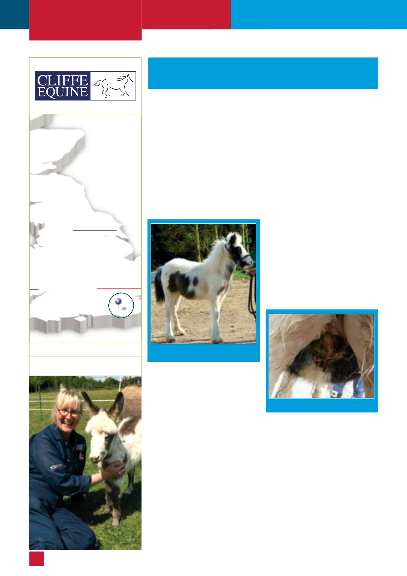
Figure 1. Bracken as a foal
Bracken, an 18 month old cob, was a very
friendly, amenable chap, having been raised
in a rescue centre from an early age
(figure
1)
. Once rehomed, his owner’s vet made
the decision to perform a standing open
castration. The procedure went smoothly
and initially the healing process proceeded
normally.
Unfortunately, a couple of weeks down the
line it became clear things were not quite
right. The owner noticed that Bracken
appeared to be walking with a slight waddle.
On closer inspection, she noticed that his
scrotum was very swollen with discharge
coming from the castration wound.
The owner rang her vet to seek further
advice. After examination, it was clear that
the castration site had become infected.
Under sedation, the incision was reopened
to ensure infected fluid was not being
retained and to allow drainage. Bracken
started on a course of broad spectrum oral
antimicrobials to try and combat infection,
along with anti-inflammatories to ease his
discomfort and reduce the swelling.
Unfortunately, Bracken’s luck was not
improving. Ten days later there was no
significant improvement in the discharge or
swelling, although Bracken behaved
completely normally otherwise. The vet
recommended further investigation and
performed an ultrasound examination of the
swollen area. This confirmed that the ends
of the spermatic cord were very thickened,
along with a lot of swelling of the surrounding
soft tissues, but no abscess had formed.
The vet recommended the best treatment for
this type of infection, known as a scirrhous
cord, was to remove the infected tissue
surgically. Correction of a scirrhous cord requires
the horse to be anaesthetised, as the horse
must be lying on his back to allow access to
the infected area. Understandably, the owner
was very worried about a second, more
invasive surgery and discussed other possible
medical options with her vet. Bracken was
given another course of oral antimicrobials,
but the infection persisted and over the next
two weeks became worse
(figure 2)
.
Feeling worried, the owner contacted the
yard from which she had rehomed Bracken
for help. The yard manager agreed to help,
and offered to care for Bracken at the rescue
centre until the problem had been resolved.
At this stage Bracken was examined by the
vets for the rescue centre, Cliffe Equine.
Working closely with the original veterinary
surgeons, all parties agreed that Bracken
needed surgery.
At surgery it was clear that about six inches
of cord was thickened and infected, but the
junction with normal cord deeper inside could
be clearly seen
(figure 3)
. Exposing and
removing a section of the normal cord was
very important to prevent recurrence of the
Figure 2. The infected castration site