


 4 / 24

3
EQUINE MATTERS
Laura Ruby BVSc MRCVS,
Calweton Equine
Veterinary surgeon
Laura Ruby
XLEquine practice
Calweton Equine
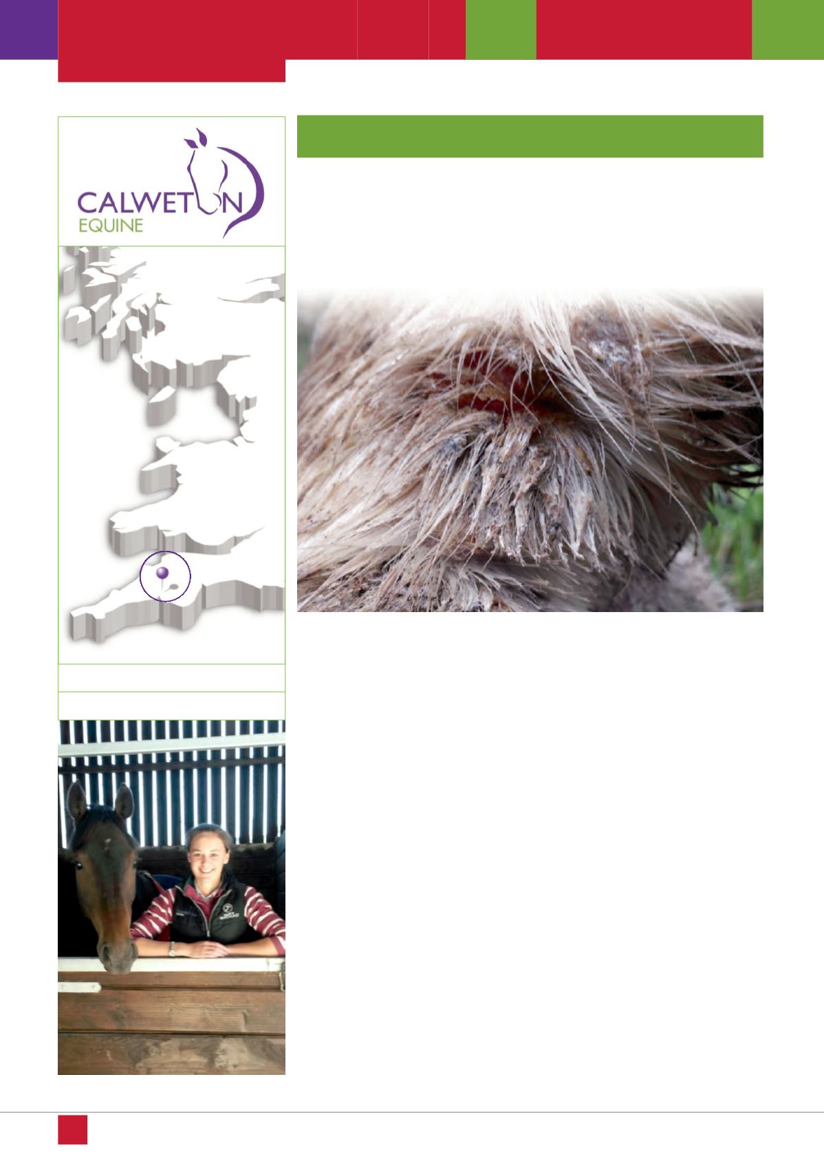
The heel bulbs and back of the pastern are
the most commonly affected areas, although
lesions can extend up the limb to the fetlock
and the back of the cannon region. Clinical
signs vary depending on the stage and
severity of infection. In the initial phase the
affected area is typically covered in multiple
small scabs. These scabs are sometimes
tightly adhered to the skin and there may or
may not be a discharge associated with
them. The skin underlying the scabs is often
very inflamed and raw-looking. The disease
process can progress if infection tracks
through into underlying tissue. In this instance
the limb becomes progressively swollen and
oedematous, is often painful to touch and a
low grade lameness may be seen.
Inflammation and infection involving the tissue
beneath the skin is known as cellulitis.
Regular inspection of the back of your horse’s
pasterns is useful for early detection of lesions.
If treatment is started as soon as a few small
scabs appear then resolution will be much
faster than if you wait until the horse is
showing significant clinical signs before
taking action.
There is some debate as to the best approach
for management and treatment of mud fever
cases. The controversy lies with whether or
not to wash affected areas. As mentioned
previously wet, damp conditions are perfect
for the bacteria to thrive. However, in the
initial phase it is important to remove the
scabs and bathe the underlying skin with an
antiseptic solution.
The following protocol is recommended for
treatment of simple cases of mud fever:
Mud fever is a common skin disease affecting the lower part of
horses’ limbs. It is most often seen during the winter period as cold,
wet weather is a predisposing factor. Ongoing wet conditions
cause the skin to soften and therefore damage to the skin barrier
occurs more easily. Mud fever is the result of infection with the
bacterium
Dermatophilus congolensis
following disruption of this
skin barrier. Once infection is established, secondary infection with
other bacteria such as
Staphylococcus spp
may occur and this can
exacerbate the problem.
Mud
fever
MU D F E V E R