


 17 / 24

AUTUMN 2015 ISSUE
LYMPHANGI T I S
Veterinary surgeon
Andrew Illing
XLEquine practice
Chapelfield Veterinary
Partnership Ltd
Lymphangitis in
horses
Lymphangitis describes inflammation of the
lymphatic vessels, and typically in horses
involves one or more legs but most
commonly a single hindleg
(Figure 1)
.
Common causes
●
Trauma
●
Allergy
●
Infection (often superficial wounds to the
lower leg)
●
Surgery
●
Genetic problems with elastic fibres in
lymphatics (more common in heavy horse
breeds especially Shires and Clydesdales)
●
Epizootic Lymphangitis (caused by a
fungal infection is not seen in the UK)
The leg receives fluid from the arterial
circulation and this fluid drains from the leg
by the veins and lymphatic vessels. The deep
lymphatic system drains most of the hindlimbs
to the pelvic lymph nodes. This is done by
smooth muscles in the lymphatic walls as
well as external forces like muscle movement,
arterial pulsation, contraction of skeletal
muscles, and joint and hoof capsule movements.
Lymphatic valves prevent back flow.
The main drainage lymph nodes are the
pre-femoral, inguinal and popliteal, but if the
lymphatics become inflamed they cannot
transport the fluid away from the leg
efficiently. The complication of inflammation
in the leg is that capillaries become leaky
and more fluid enters the tissues. Quickly the
system becomes overloaded and the leg gets
rapidly bigger.
Clinical signs
The leg can have a mild form of fluid
accumulation ‘lymph-oedema’, where fluid
passively collects because of gravity at the
bottom of the leg. This is passive with no
inflammation and is non-painful. This is often
a result of long periods of stabling, weight
gain or pregnancy. This resolves with light
exercise and bandaging.
Lymphangitis is:
●
Very painful (the leg may not be able to bear
weight)
●
The leg is very hot and very swollen (usually
a hindleg, but can be a front leg, or more
than one)
●
The leg may feel doughy (pitting oedema),
but painful to touch
●
Often the horse is running a temperature,
with increased respiratory and heart rates
Where there are skin lesions of the pastern
(mud fever), or small abrasions of the skin, such
as Chorioptic mange
(Figure 2)
aggravation,
secondary bacterial infection with
Staphylococcus or Streptococcus
species may
lead to a cellulitis infection spreading up the leg.
This may cause a secondary lymphangitis with
the leg developing ulcers or starting to weep
serum
(Figure 3)
.
Diagnosis
Ultrasound or radiographs may be used to rule
out fractures or other soft tissue injuries which
sometimes give similar symptoms. Confirmation of
infection is not always straightforward but swabs
may be taken if the skin is weeping serum.
Treatment
●
Antibiotics – trimethoprim/sulphadiazine or
penicillins; usually a long course of
medication is needed
●
Non-steroidal anti-inflammatories –
(phenylbutazone, flunixin, meloxicam)
reduce inflammation and help to bring the
temperature down
●
Corticosteroids – Dexamethasone (may
not be given if there is a specific risk
of laminitis)
●
Diuretics (remove fluid from the body)
●
Frequent gentle exercise and physiotherapy
●
Bandaging after having an antiseptic wash
if weeping serum
Treatment will often leave the horse with a
larger leg, prone to recurrence, due to
damage to the lymphatics and the
subcutaneous tissues.
Andrew Illing VetMB MA MRCVS,
Chapelfield Veterinary Partnership Ltd
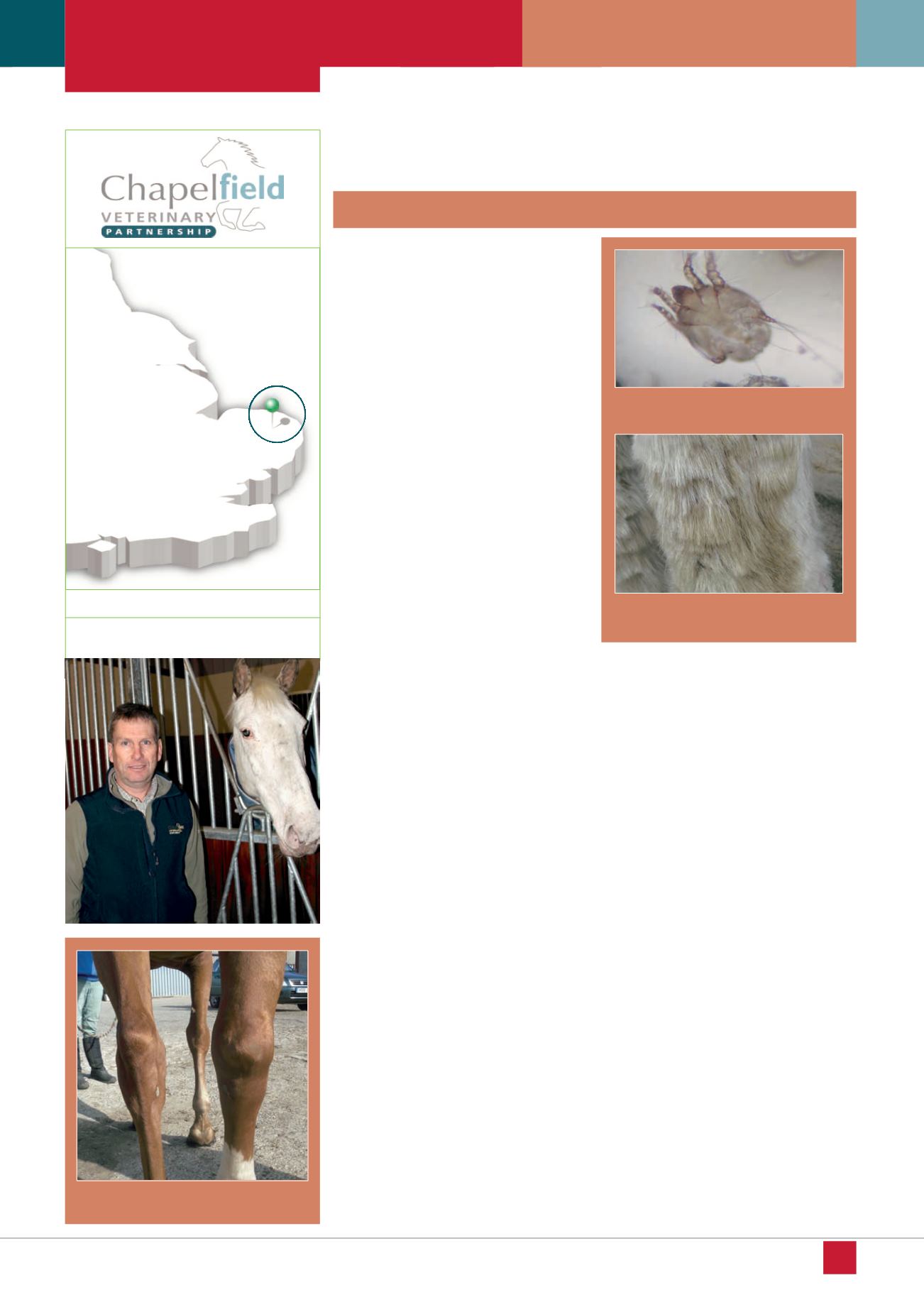
Figure 1 – Lymphangitis affecting a single
hindlimb
Figure 2 – Chorioptes mites can cause
abrasions in the skin
Figure 3 – Serum weeping from superficial
wounds
EQUINE MATTERS
16