


 17 / 24

AUTUMN/WINTER 2016 ISSUE
EQUINE MATTERS
16
Figure 1. Tail bandages and travel boots
help to reduce injury during travel
Figure 2. Horses should be unloaded at least
every four hours on long journeys
I was asked to examine ‘Bob’, a 21 year old Friesian gelding,
because he wasn't eating very well. His owner was worried that
he was losing weight and wasn't eating hay as quickly as usual.
By observing ‘Bob’ eating, I noticed he was reluctant to pull
hay from the hay net, preferring to pull his hay downwards to the
floor before eating it slowly.
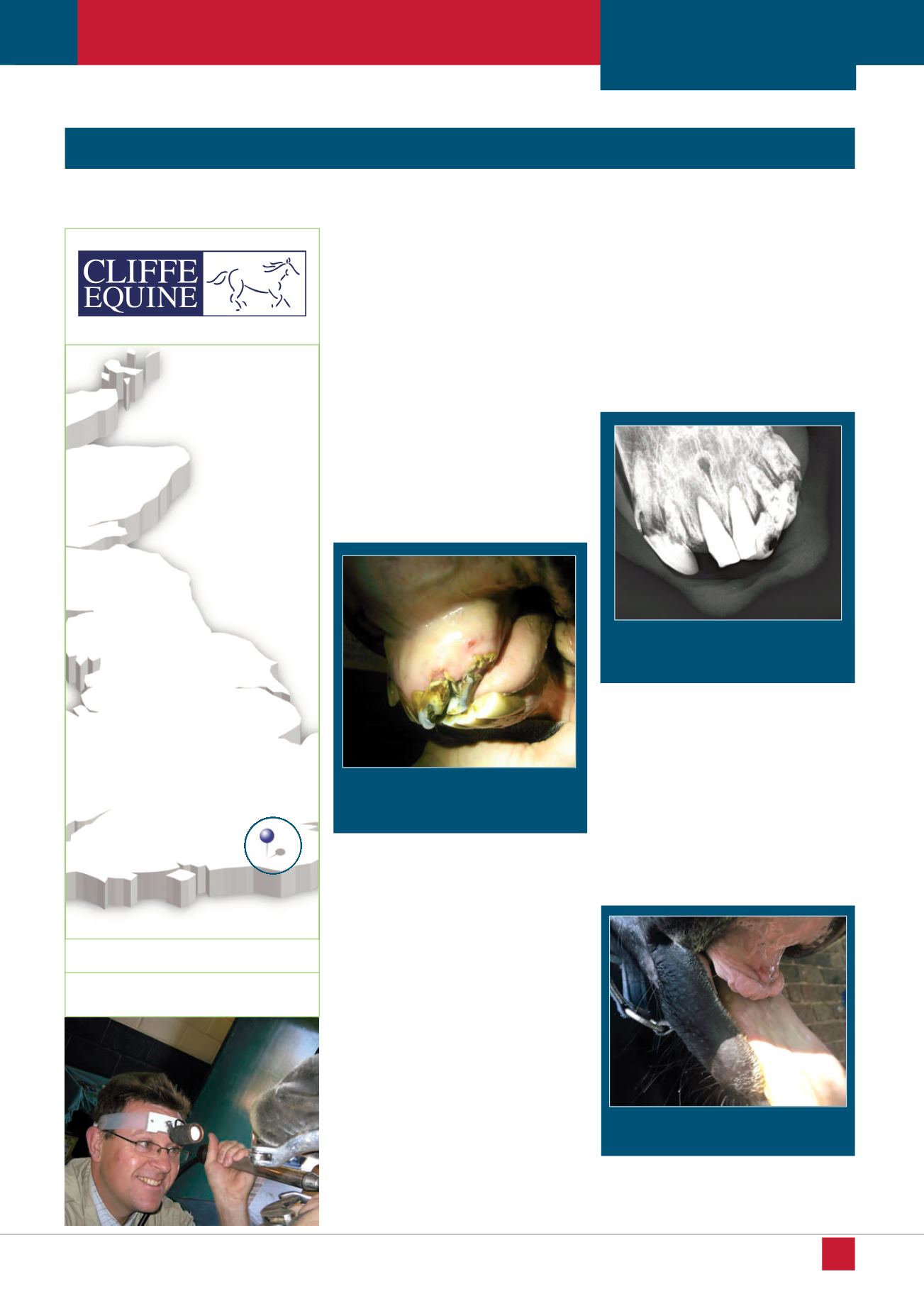
Figure two. This x-ray shows the broken
incisors and changes to the tooth root
structure
Figure three. Bob’s gummy, but pain free
smile!
Surgical treatment of a common aged horse disease
Dr Karl Holliman,
BVM&S CertEP MRCVS, Cliffe Veterinary Group
FOCUS ON DENTISTRY:
EOTRH
Veterinary Surgeon
Karl Holliman
XLVets Equine practice
Cliffe Veterinary
Group
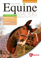
Figure one. View of Bob’s incisors from
the left side, showing the tooth angulation
and altered structure
The mouth was examined first by palpating the
head, then by parting the lips to examine the
incisors (front teeth) and canine teeth. The
incisors were in a poor state, with upper teeth
displaced at angles and disruption of the
surface with loss of the normal smooth
appearance
(figure one)
. The gums had some
reddening around the gingival margin with a
build up of cement. The jaw bone supporting
these teeth was also swollen.
Heavy sedation was required to allow
examination of his cheek teeth; even then the
placement of the gag, which has plates that sit
on the front incisor teeth allowing the mouth to
be held open, was very painful for ‘Bob’.
Fortunately, there were no significant dental
abnormalities of the molars and premolars,
so I concluded the pain originated from the
pressure on the front teeth.
This was a classic case of Equine
Odontoclastic Tooth Resorption and
Hypercementosis, more simply known as
EOTRH. EOTRH is not an uncommon disease
of older horses that affects the incisors. This
disease is poorly understood, but is very
destructive, leading to the breakdown of the
tooth enamel with severe inflammation and
reaction around the roots of these teeth. Any
pressure, such as chewing or gag placement,
can lead to marked pain.
The incisors were radiographed
(figure two)
w ich rev aled the destr ction of the dental
tissues below the gum line and the increase in
cementum around the tooth roots, confirming
the severity of the disease.
Under heavy sedation and local anaesthesia
(nerve blocks), all the upper incisors and
several of the more diseased lower teeth were
extracted. Radiographs were taken to confirm
removal of all dental tissue. The tooth sockets
were flushed and packed with antiseptic
material and ‘Bob’ went home later that day.
When I visited ‘Bob’ again three days later it
was wonderful to see him eating happily and
with enthusiasm from his hay net! With daily
flushing, the gums healed over a four week
period without complication.
Over the next six months, ‘Bob’ quickly
return d to a h alt y weight and th own r
reports that he grazes and eats well, despite
the lack of front teeth
(figure three)
!