Basic HTML Version


SARCOI DS
Richard Morris BSc BVetMed CertVD MRCVS
Fenwold Veterinary Practice
CASE REPORT...
Surgical Feature:
Equine Sarcoids
Equine sarcoids are the most common equine
tumour accounting for over half of all equine
tumours. They are thought to be triggered by
infection with the Bovine Papilloma virus BPV1
and 2. Their behaviour is unpredictable and
inactive sarcoids may become aggressive if
disrupted by injury, surgery or inappropriate
treatment. There are many approaches to
management and the size and number of
sarcoids will determine the technique
for removal.
Surgical sarcoid removal involves physically
removing the sarcoid and can include ligation,
conventional excision and laser surgery.
We shall discuss the surgical treatment of
sarcoids and illustrate these techniques with
case studies.
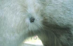
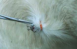
Ligation with a rubber band or tying a ligature
with suture material around isolated individual
sarcoids can be carried out in a select number
of cases but the site and type of sarcoid has
to be appropriate; they need to be easily
accessible with plenty of loose skin. The
verrucose sarcoid in figures 1 and 2
responded well to this treatment. The horse
may be restrained with a twitch or sedation
may be required. The sarcoid could be
treated with cryotherapy at the same time
to improve effectiveness.
More extensive sarcoids may need removing
with conventional surgery which can be
carried out under standing sedation and local
anaesthesia or in some cases may require
general anaesthesia. This will depend on the
size and location of the sarcoid. It is necessary
to remove a 2-3cm margin of healthy tissue
around the edge of the sarcoid (which may
contain seeds of the original tumour) in order
to prevent recurrence. Success rates of
30-50% are quoted with conventional
excision, most relapses occurring within about
six months, sometimes in a more aggressive
form so 'regrowths' should be treated as soon
as they appear.
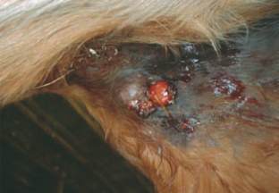
Figure 3 shows a nodular sarcoid in the groin
of a chestnut mare removed under standing
sedation and local anaesthetic. In this case,
no relapse was seen after five years. Skin is
elastic and contracts once the sarcoid is
excised so wound edges appear smaller than
the removed sarcoid.
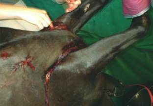
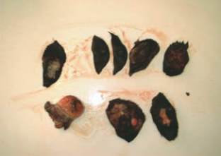
Figures 4 and 5 show extensive sarcoids
requiring general anaesthesia. The sarcoids
had previously been treated with the Liverpool
sarcoid cream AW3-Ludes but they re-grew
soon after treatment. The decision was made
to remove every sarcoid visible on the horse at
that time under a general anaesthetic. The
horse made a full recovery and no relapses
were seen nine years later.
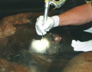
Surgical removal with a laser uses carbon
dioxide to cut and vaporise the tissue around
the sarcoid (figure 6). This causes less pain
and swelling and minimal bleeding compared
with conventional surgery with success rates of
60-80% reported.
Complete resection of sarcoids can be difficult
so other treatment options may be used
alongside surgery including applying topical
ointments after surgery such as Imiquimod or
Aciclovir. Sarcoids are a major therapeutic
challenge; early recognition and treatment
reduces the complications and improves the
outcome when treating these tumours.
Veterinary Surgeon
Richard Morris
XLEquine Practice
Fenwold Veterinary
Practice
EQUINE MATTERS
12
WINTER 2014 ISSUE
Figure 4:
Removal by conventional excision
Figure 5:
Extensive sarcoids after removal
Figure 6:
Surgical removal of sarcoids with a
laser using carbon dioxide
Figure 1:
Verrucose sarcoid
Figure 2:
Ligation with a rubber band
Figure 3:
Nodular sarcoid in the groin of a
chestnut mare