Basic HTML Version


When examined he had a thick nasal
discharge and swollen glands (lymph nodes).
His temperature was 41°C but he was
otherwise normal. A new horse had recently
been brought onto the yard with a mild nasal
discharge but was otherwise healthy. Strangles
was immediately suspected and so a
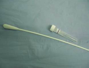
nasopharyngeal swab (Figure1) was taken
from Bob and the new horse, to confirm the
disease. The swab was passed through the
nostril close to the back of their throats and
they were made to swallow. The livery yard
owner was advised on the likelihood of a
strangles outbreak and so the yard was closed
immediately and a strict biosecurity regime
was instigated. Results from both swabs
confirmed strangles 48 hours later.
Strangles is a bacterial respiratory infection
caused by Streptococcus equi. It causes
abscesses to develop primarily in lymph nodes
and is spread by direct contact with nasal
discharge (nose to nose touching of horses or
on contaminated water buckets, clothing and
tack etc) and is, uncommonly, fatal.
Bob was treated with anti-inflammatories
for three weeks. No antibiotics were given as
is often the case when abscesses have
developed; these were encouraged to burst
by hot compressing.
Other owners on the yard were advised
to take their own horse's temperatures twice
daily. If a temperature was noted above
normal (38.5°C), antibiotics were
administered immediately. A high temperature
is the first clinical sign of strangles; if observed
promptly antibiotics can be administered
before abscesses develop. Any horse that
developed a high temperature was moved
into group 1.
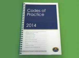
Horserace Betting and Levy Board (HBLB)
codes of practice advise Veterinary Surgeons
(Figure 2) that three negative nasopharyngeal
swabs taken one week apart, or one negative
guttural pouch wash are necessary from
every horse in group one before the yard can
be opened. Tests began six weeks after the
last horse in group one developed a
temperature. At this time all horses including
Bob appeared healthy.
A guttural pouch wash is taken by passing
an endoscope into the guttural pouch via the
horse’s nose. It is an important space, housing
major vessels and nerves where the bacteria
can 'hide' in low numbers. Saline was flushed
into and drawn back out of both the left and
right pouches of all horses in group one,
followed by an infusion of penicillin. Samples
were then tested for Streptococcus equi.
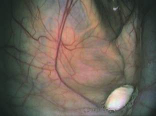
Endoscopy of Bob's left guttural pouch revealed
chondroids (Figure 3), these are dried pus-like
structures that contain low numbers of bacteria.
The chondroids were eventually 'dissolved'
and removed by repeatedly infusing a
mucolytic drug into the guttural pouch. A final
wash was taken from Bob 4 months after he
was diagnosed with strangles and declared
he was negative. All other horses on the yard
were negative at the initial guttural pouch
wash. Bob was the worst affected by the
disease and took the longest to recover.
However, the following year he was back in
the show ring with no signs that he had been
so ill the previous year!
STRANGL ES
Veterinary surgeon
Gemma Dransfield
XLVets Equine practice
Minster Veterinary
Practice
CASE REPORT...
Gemma Dransfield MA VetMB CertEP MRCVS
Minster Veterinary Practice
Case Study:
Strangles
'Bob'
a four year old cob
was presented to the practice
as he was not eating and had
a snotty nose.
Figure 2:
The HBLB codes of practice that
Veterinary Surgeons follow when treating a
Strangles outbreak on a yard.
Figure 3:
An endoscopic view in a horse's
guttural pouch. The large white mass is
a chondroid.
Figure 1:
A nasopharyngeal swab and
bacterial growth media required to diagnose
Strangles infection.
15
EQUINE MATTERS
1.
infected horses, such as Bob;
2.
in-contact, horses that had been in
contact with horses in group one;
3.
non-contact, horses that had not been
in contact with horses in group one.
All horses on the yard were divided
into three groups: