Basic HTML Version


SUMMER 2013 ISSUE
Put simply, the assessment of foot balance
is generally carried out initially by visual
inspection in the resting, weight bearing
position on a firm level surface from the side,
from in front and from behind. The foot can
also be viewed from above when it is picked
up and, with the sole perpendicular to the
ground, the side to side balance or
medio-lateral (M-L) balance can be assessed.
The use of an instrument called a T-square
can also help gauge any imbalance.
A side view of the foot is aiming to assess the
alignment of the hoof wall at the toe, relative
to the hoof wall at the heel and to the angle
of the pastern bones up to the fetlock joint.
This is termed the hoof pastern axis (HPA).
The normal acceptable range of angle is
roughly 45-50° for forelimbs and 50-55° for
hindlimbs, with some breeds such as the TB
being slightly less, while a Cob for example
might be expected to have a more upright
angle. Ideally, a straight line should be able
to be drawn from the toe, along the front of
the pastern to the fetlock (Figure 1).
A horse with an upright or broken forward
HPA would have a shallower pastern angle
compared with the front of the hoof wall and
would tend to occur in animals with boxy or
clubbed feet (Figure 2). In contrast, a horse
with a broken back HPA would have a steeper
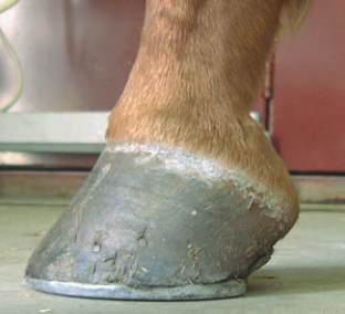
pastern angle (Figure 3) and can be seen in
animals with long toes and collapsed heels.
Both these abnormalities can predispose to
injury and associated lameness if uncorrected.
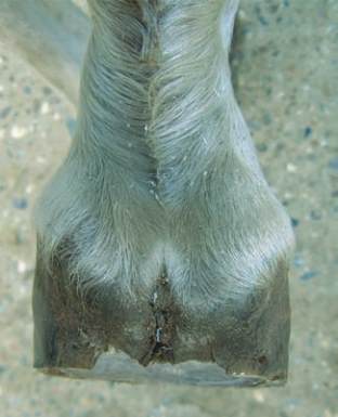
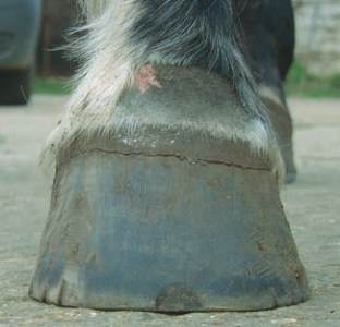
When viewed from in front, behind or above
(Figure 4) with the foot elevated, the M-L
balance can be assessed. The M-L foot shape
should normally be more or less symmetrical,
although many horses are not perfect. M-L
imbalance can result in the hoof wall and
coronet being higher on one side than the
other (Figure 5), or the quarter/toe wall
growing out at a different angle from the other
side, causing the foot to ‘drift’ inwards or
outwards. Any M-L imbalance will become
self-perpetuating as the hoof wall will tend to
grow in the direction of the imbalance. The
consequences of M-L imbalance are an
alteration in the distribution of weight bearing
forces passing through the foot and limb,
resulting in tissue injury and lameness within
the foot and further up the limb.
Veterinary Surgeon
Chris Lehrbach
XLVets Equine Practice
Chapelfield Veterinary
Partnership
Foot balance explained
Whilst advances in science have
resulted in the occasional reports
of equine prosthetic lower limbs,
which would undoubtedly have
saved the patient's life, without a
foot there is still generally no
horse. One particularly problematic
aspect of the horse's foot is its
shape, a feature of its conformation
termed the foot balance.
FOOT BALANCE
Chris Lehrbach BVMS MVM Cert ES(Orth) MRCVS,
Chapelfield Veterinary Partnership, Brooke Equine Clinic
Figure 3.
A broken back hoof pastern axis,
with upright pastern compare to hoof wall
Figure 2.
A broken forward hoof pastern
axis, with upright hoof wall
Figure 1.
A normal hoof pastern axis, the
front of the hoof wall and pastern area
being aligned
EQUINE MATTERS
12
Figure 4.
Side to side foot imbalance
viewed from above, with asymmetry of
the hoof wall
Figure 5.
Side to side foot imbalance
viewed from in front, with one side of the
hoof wall higher than the other