Basic HTML Version


The images are obtained by placing the area
to be examined in a magnetic field. This area
is then bombarded by a series of pulses of
radiowaves. The signal is received back by a
coil within the machine which converts them
into a series of 3D images. The procedure is
low risk to the patient and handlers because
ionising radiation (used in radiography and
bone scans) is not used.
Types of MRI imaging
MRI can be 'high field' using very high magnet
strength, which produces the most detailed
images; however the horses need to be given
a general anaesthetic to go into these machines.
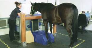
The majority of equine MRI imaging done in
the UK is 'low field' (using a weaker magnet)
but the units are designed for horses to be able
to walk in and be examined under sedation.
The indications for an
MRI examination
Only small areas can be examined for an MRI
study so it is not realistic to obtain images of
a whole leg. It is therefore important that the
lameness has been clearly isolated to the foot
region using nerve or joint blocks and that a
standard set of x-rays have been completed
prior to considering MRI imaging. In cases
where changes in the bones or coffin joint are
involved, a diagnosis may be made following
x-ray examination. MRI is a useful tool for
further investigation where significant x-ray
changes are not present or if the horse remains
lame despite initial medical therapy.
MRI is a tool for diagnosis, not for treatment,
and not every horse with foot pain needs an
MRI. It will however help to give an accurate
diagnosis of what structures are involved in
causing a horse foot pain in cases where
further imaging is indicated. A definitive
diagnosis then allows your vet to discuss
available treatment options and the likely
prognosis of a return to soundness and
function for your horse's condition.
Veterinary Surgeon
Wendy Furness
XLVets Equine Practice
Scarsdale Veterinary
Group
MRI
of the equine foot
MRI (magnetic resonance imaging) i
s an advanced imaging technique
that produces highly detailed cross sectional anatomical images of
bone and soft tissue structures in the area being examined.
MR I IMAGING
Wendy Furness MA VetMB CertEP MRCVS,
Scarsdale Veterinary Group
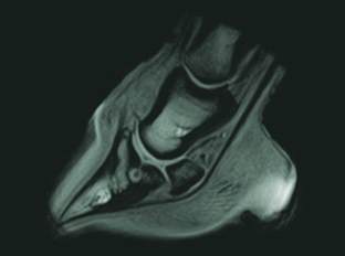
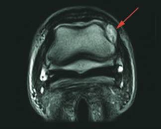
MRI image showing a bone cyst within the
pedal bone
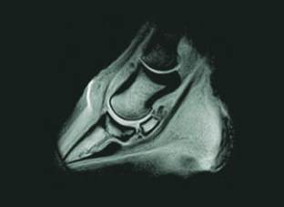
MRI image showing fragments on the border
of the navicular bone
Transverse MRI image showing damage to
the collateral ligament of the coffin joint
The advantages of MRI
The images obtained have excellent detail
so offer a lot more information than
conventional imaging techniques such as
radiography and ultrasound. Ultrasound
waves cannot pass through the hoof
capsule so our ability to obtain good soft
tissue images of the foot using ultrasound
is very limited.
MRI
allows assessment of the bones and
cartilages within the foot and can be useful
in diagnosing the following foot conditions:
l
changes to the navicular bone structure;
l
damage to the deep digital flexor tendon;
l
damage to the small supportive ligaments
of the navicular bone;
l
injuries to the collateral ligaments of the
distal interphalangeal joint (coffin joint);
l
changes to the joints including the distal
interphalangeal joint (coffin joint) and the
proximal interphalangeal joint (pasternjoint).
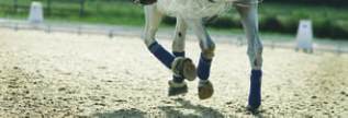
Horse standing in MRI
11
EQUINE MATTERS