Basic HTML Version


SPRING 2012 ISSUE
EQUINE MATTERS
8
A R T H R I T I S
Case History
Midnight was a ten-year-old cob cross mare
used for general pleasure work including
riding club activities. The day following
cross-country schooling she was found lame
on the left hindlimb with a large swelling at
the back of the fetlock. Midnight was box
rested, given anti-inflammatory therapy (oral
phenylbutazone (Bute) and hosing of the
area but remained lame over the next
10 days. This prompted the vet to refer the
mare to Clyde Vet Group Equine Hospital
for further investigations.
Assessment
Midnight was 5/10ths lame and had a
swollen, hot and painful left hind windgall.
X-rays did not demonstrate any bony damage
but ultrasound scans revealed considerable
thickening of the sheath lining and the
suspicion of damage to the outer (lateral)
side of the tendons.
Surgery
Due to the degree of lameness and the
possible tendon damage it was decided to
take Midnight to surgery for keyhole assessment
of the sheath. General anaesthesia was
induced in a padded knockdown room before
Midnight was moved to the operating room
and positioned on her right side so the affected
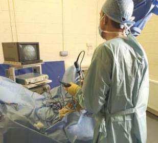
windgall was uppermost. After sterile
preparation, an arthroscope (a 4mm diameter
telescope connected to a camera) was inserted
into the sheath just below the sesamoid bones
(Figure 2). From this site it is possible to
visualise the entire contents of the sheath.
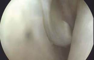
Examination revealed a tear to the outside of
the manica flexoria portion of the superficial
digital flexor tendon (Figure 3). Another small
incision was made in the upper portion of the
sheath through which a variety of instruments
were placed. Using these it was found that the
torn portion of the manica was adhered to the
inside of the sheath.
A small motorised resector (3mm diameter)
was then used to break down the adhesions
and remove the torn tissue. The resector has
a small rotating blade that oscillates at high
speed whilst at the same time it removes all
the resultant debris. Finally the sheath was
washed out with saline before the skin
incisions were closed and a heavy support
bandage placed on the limb. Midnight then
returned to the recovery room to wake up
from anaesthesia; she was helped to get to
her feet by a system of ropes and pulleys.
Over the next five days she received
antibiotics and anti-inflammatory drugs
before returning home. The limb remained
bandaged for two weeks.
Midnight was stabled for six weeks
before being turned out for three months.
Re-assessment then revealed her to be sound
and there was only a minor degree of sheath
distension. Repeat ultrasound scan revealed
no abnormalities and it was recommended
that she should resume normal work.
Two years following the surgery the owner
reports Midnight to be sound and she is
being used for riding club work.
Discussion
Tears to the manica flexoria are increasingly
recognised as a cause of lameness in cob type
horses. They often require surgery for correction.
If your horse does develop a large windgall
please contact your veterinary surgeon.
Case Study:
Lameness - surgical treatment
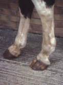
Figure 1
Appearance of
a distended tendonous
windgall.
Figure 2
Keyhole surgery conducted on a
horse's stifle joint demonstrating how the
surgeon uses the monitor to assess the interior
of joints or tendon sheaths.
Figure 3
Appearance of the torn manica
flexoria seen through the camera placed in
the windgall. The torn manica is in the centre
of the screen and was found to be adhered
to the lining of the sheath behind it. The
adhesions were broken down and the torn
portion removed guided by the scope.
A troublesome windgall requiring surgery
Andrew McDiarmid BVM&S CertES(Orth) MRCVS
, Clyde Veterinary Group
V
ets commonly use the term windgall to describe fluid swellings in the fetlock area. There are
two types of windgalls, tendonous and articular windgalls. The more common tendonous
windgalls form due to increased fluid within the tendon sheath surrounding the flexor tendons
as they traverse over the back of the fetlock (see Figure 1). This report is of a horse with a
tendonous windgall that required keyhole surgery.
Veterinary Surgeon
Andrew McDiarmid
XLVets Equine Practice
Clyde Veterinary Group
1
2
3