Basic HTML Version


L AM E N E S S
SPRING 2012 ISSUE
EQUINE MATTERS
4
Figure 3
The horse is walked and trotted on
a firm surface
3. Gait analysis
The next part of the lameness examination
will evaluate the horse's gait. The horse will
be observed at walk and trot in-hand (see
Figure 3). The vet may require your horse to
be walked or trotted in-hand, lunged (Figure
4), or ridden on soft and hard ground.
Usually at this stage of the examination it
will become apparent if the horse is lame
and if so, which leg/legs are affected.
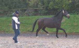
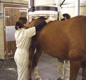
Figure 4
Lunging is often used in the
assessment of lameness
4. Flexion testing
Flexion tests may be helpful if the lameness
is subtle or there are no obvious signs of a
problem. Typically, flexion tests involve
bending or "flexing" a joint for 30 seconds
to two minutes. Then the horse is trotted for
about 20-30 metres and evaluated for an
increase in lameness. If a particular flexion
test intensifies the lameness, your vet may
concentrate on that area of the body or
that joint as the source of the lameness
during the rest of the examination.
When performing diagnostic nerve blocks
(shown in Figure 5), local anaesthetic is
infused either around a nerve or injected
into a joint or other synovial structure,
e.g. tendon sheath, bursa, etc. If the
lameness disappears or improves markedly
following administration of anaesthesia,
your vet will have successfully localised
the site of lameness.
After nerve blocks have localised an area
causing the lameness, diagnostic imaging,
e.g. radiography, ultrasonography, etc,
can be used to further understand the
lameness condition. If the lameness
cannot be localised by nerve blocks,
other diagnostic imaging techniques can
be employed. Occasionally, the process of
‘blocking’ will be by-passed and diagnostic
imaging will be performed, particularly if
the affected region is already identified.
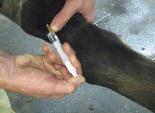
Figure 5
A nerve block being performed
5. Diagnostic local anaesthesia
(nerve or joint blocks)
6. Diagnostic imaging
Radiographs (x-rays) are used to show
changes in bone and joint surfaces once
the area causing the lameness has been
identified. X-rays can be extremely useful
in the investigation of lameness but require
careful interpretation however, as x-rays
may reveal historical changes in the
bone which are not related to the current
lameness. They are of limited usefulness in
the investigation of soft tissue injuries.
Magnetic resonance imaging (MRI) and
computed tomography (CT): Magnetic
resonance imaging is revolutionising the
field of equine diagnostic imaging and
orthopaedics. MRI scans use magnetic fields
to create images of the tissues inside the
body. MRI scans are increasingly common
in the investigation of lower limb and foot
conditions. CT scans use multiple X-rays to
create three-dimensional images of an area
and can provide high quality images of
both bone and soft tissue structures.
Figure 6
Radiograph of a horse suffering
from chronic laminitis
Figure 7
Ultrasound image of tendons in
a horse
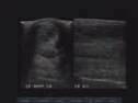
Ultrasonography (‘ultrasound’) is the most
practical and accurate method for
assessing tendons (see Figure 7),
ligaments, and other soft tissues. Ultrasound
is an excellent way to view the soft tissue
structures in the horse. It allows real-time
imaging of the tendons and ligaments.
Figure 8
A bone scan of the pelvis being
performed in a horse
6
7
Scintigraphy (bone scan) is usually
performed in horses when the lameness
is very subtle, intermittent, or when a
non-displaced fracture (small crack) is
suspected. Bone scans are useful in horses
which are not amenable to nerve or
joint blocks.
During scintigraphy, the horse is given
an injection, which contains a small
quantity of radioactive material. The
radioactive material will become
distributed throughout the horse’s body
and can be detected using a gamma
camera, which is linked to a computer.
The information gained can be used to
determine sites of abnormality and thus,
possible causes of lameness. Scintigraphy
is safe for horses, but the horse must
remain at a special quarantine facility
for a short period following injection
of the radioactive material, usually for
24 - 48 hours.
The investigation of the lame horse involves
a structured and careful approach from
your vet which will ensure the highest
chance of finding the cause of the lameness.
The actual approach used by your vet may
utilise some, or a combination of these
techniques in order to discover the cause
of lameness.
IN SUMMARY
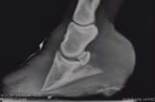
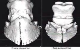
Figure 9
CT Scan: a 3-Dimensional image
of a fractured pedal bone