Basic HTML Version


7
EQUINE MATTERS
SUSPENSORY LIGAMENT
Suspensory ligament
disease
in the horse
Veterinary Surgeon
Louise Cornish
XLVets Equine Practice
Clyde Veterinary
Group
Louise Cornish BVMS CertEP MRCVS,
Clyde Veterinary Group
The suspensory ligament in the horse supports the fetlock and is very prone
to injury in many breeds and ages of horses. Inflammation of the ligament
(desmitis) is increasingly recognised as a cause of lameness in sports horses
especially. This article aims to give some background information regarding
the anatomy of the ligament, the areas which are susceptible to damage,
how the condition can be diagnosed and what treatment is available.
Anatomy
The suspensory ligament originates at the top
of the back of the cannon bone, just under
the knee or the hock, travels downwards
between the splint bones and then divides
into two branches to attach onto the sesamoid
bones at the back of the fetlock joint. The
origin of the suspensory ligament is also
known as the proximal suspensory ligament.
The middle part (the body) also contains some
muscle tissue which can become stronger with
training. The two branches which join onto
the fetlock can also be injured.
Smaller, less significant extensor branches then
travel to the front of the pastern where they join
the extensor tendon. These are rarely damaged
and will not be discussed further here. Several
short ligaments run from the base of the
sesamoid bones (at the back of the fetlock
joint) down the back of the pastern. These are
part of the suspensory apparatus and can be
a source of lameness.
Like all ligaments and tendons in the body, the
suspensory apparatus is subject to overload
and strain. It acts as a sling for the fetlock in
the fore and hind limbs. It is more likely to be
injured in older horses and in horses with a
straight hindlimb conformation. Often both
limbs are affected.
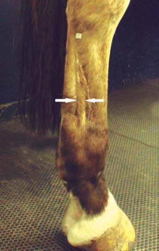
The suspensory ligament can easily be seen
and felt in the mid cannon region (between
the arrows)