Basic HTML Version


CO R N E A L D I S E A S E
Equine corneal
disease
Veterinary Surgeon
Dominic Alexander
XLVets Equine Practice
Belmont Veterinary
Centre
Dominic Alexander BSc BVMS MRCVS,
Belmont Veterinary Centre
The eyes on all wild and domestic horses and donkeys are large and placed
on the corners of the head giving them almost a 360 degree field of vision.
The size and prominence of the equine eye means that disease and injury to
the equine cornea is a common occurrence. Many incidences are minor and
heal before they come to our attention.
What is the cornea?
The cornea is the transparent front part of the
eye that covers the iris, pupil and anterior
(front) chamber. The anterior chamber is the
fluid-filled space inside the eye between the
lens and iris, and the inner surface of the
cornea. The transparent nature of the cornea,
and the lens, allows light to pass through the
eye to the back of the eye.
The healthy cornea is surprisingly robust
considering that it is less than one millimetre
in thickness. This thin structure comprises of
several layers. The outer membranes of the
cornea are highly innervated with nerve
fibres which makes any damage to the
cornea very painful.
Assessment of the eye
Occasionally dust or any sort of debris can
get into the eye and cause irritation. Excess
tear production, a mucky discharge or even
some squinting of the eyelids may be seen.
Wiping the eye with a piece of kitchen
towel soaked in warm water may be all that
is required to remedy the problem. Using
proprietary human eye washes can help
soothe the eye. Salt water or cold tea, are
sometimes used but they can dehydrate/dry
out the cornea if strong concentrations are
used doing more harm than good.
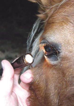
Figure 2
- much of the front (anterior) part
of the eye, including the cornea, can be
examined with a small torch
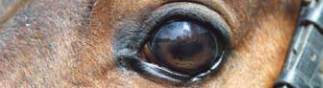
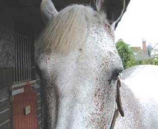
Figure 1
- note the eyelash on the affected
left eye (red arrow) is angled downwards in
comparison with the eyelash on the normal/
healthy right eyelid (yellow arrow)
●
squinting/closing of the eyelids
●
excess tear production
●
sticky cream/grey discharge in the
corner of the eye
●
reddening around the edges of
the eye
●
increased sensitivity around the eye.
●
loss of the smooth shiny finish of the
cornea (this is particularly noticeable
when the eye is 'back lit' (see Figure
2) using a small torch with the beam
directed towards you)
Corneal disease
- signs to
look out for:
●
rubbing the eye
11
EQUINE MATTERS