Basic HTML Version


A I RWAY ANAT OMY
15
EQUINE MATTERS
Veterinary Surgeon
Mark Tabachnick
XLVets Practice
Wright & Morten
Macclesfield
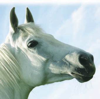
The horse's airway is divided into the upper respiratory
tract; which starts at the nostrils and ends at the larynx
at the back of the throat, and the lower respiratory tract;
the trachea and the lungs.
Airway Anatomy
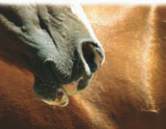
The nostrils
Horses’ nostrils are naturally very large, but
are also very flexible. They are supported by
a cartilage called the alar cartlilage, and
have a well developed muscle attachment.
This means at strenuous exercise, they are
capable of massive dilation to allow in more
air. Within the nostril there is a blind ending
pouch called the false nostril, which appears
to have no anatomical function.
The nasal cavity
The nasal cavities stretch from the nostrils to
the throat. They are divided into a series of
narrow passageways by thin strips of bones
called turbinates. In between the turbinates
are a series of passageways where the air
runs freely. The turbinates are lined by a layer
of tissue with a good blood supply called
mucosa. This functions to warm and moisten
the air before it reaches the lungs.
The nasal cavities overlie the tooth roots of
the horse's molar teeth, and are closely
connected to the sinuses. The sinuses are a
series of air filled chambers within the horse's
skull. Their exact function is unknown. They
may have evolved to allow the bony skull to
be relatively light. The sinuses communicate
with the nasal cavity via a small opening.
At the back of the nasal passages are a
number of mushroom shaped projections
called
ethmoturbinates
. These are important
for the horse's sense of smell.
Mark Tabachnick BVM&S, BSc, CertEP, MRCVS