Basic HTML Version


WOUND G R A F T I NG
5
EQUINE MATTERS
Skin Graft
Case Study:
Veterinary Surgeon
Louise Cornish
XLVets Practice
Clyde Veterinary Group
Oliver had unfortunately fallen for the old
saying ‘the grass is always greener on the
other side of the fence’ and had decided to
explore when he caught his left hind limb in
a plain wire fence. He was found limping
in the neighbouring field by his distraught
owner, who discovered that he had a large
wound to the front of the leg from above
the hock to the upper part of his cannon
bone. The wound was deep in places,
narrowly missing the hock joint itself but
scraping the surface of the cannon bone.
Most of the skin in the area appeared to
be missing, and what was left was too
damaged to be stitched together.
The vet was called and administered
painkillers, antibiotics and a sedative. The
wound was cleaned, taking as much dead
tissue away as possible. Oliver was stabled
with a well-padded bandage to protect the
wound and to reduce limb movement. He
became much more comfortable over the
next few days but was kept on box rest,
despite his protestations, to keep movement
to a minimum which would otherwise slow
down wound healing.
In this type of wound, healing occurs by
‘second intention’, which means that there is
a gap between the two edges of the wound
which must initially be filled by granulation
tissue. The edges of the healthy skin then
begin to develop new skin cells and
gradually grow, cell by cell, over the
surface of the bed of granulation tissue. If
the granulation tissue grows higher than the
surface of the normal skin, it is termed
proud, and needs to be removed surgically
or by applying creams or powders;
otherwise it will interfere with the new
skin growth. A firm padded bandage
will effectively help to prevent proud
flesh development.
Although Oliver’s wound initially appeared
to be healing well, progress seemed to
cease after about 6 weeks. Proud flesh
developed and the wound edges were no
longer producing new growth of skin cells.
It was decided to perform a skin graft to
speed up healing. Under sedation and
local anaesthesia, about 20 small sections
of skin were removed from Oliver’s neck
using a punch biopsy instrument. These were
embedded into the granulation tissue in the
hock region and kept in place by bandaging.
The graft provided a large number of
healthy skin cells to multiply and help to
cover the wound with new skin. Within a
fortnight, the skin graft had adhered well to
the granulation tissue bed and healing was
again progressing nicely. A further month
later, the wound was small enough to leave
open although box rest was continued. A
prescription gel was applied twice daily to
prevent proud flesh growth.
Four months after the initial injury, Oliver’s
wound had fully healed, leaving only a scar
and he was able to rejoin his friends in the
field for some early spring grass!
W
ounds on the lower limbs of horses are very common but notoriously slow to heal and frequently
develop ‘proud flesh’. This case report follows the progress of Oliver, an 8 year old Arab gelding,
who injured his leg when caught in a wire fence. A skin graft was used to aid healing.
Louise Cornish BVMS Cert EP MRCVS,
Clyde Veterinary Group
Use of a skin graft in a limb wound
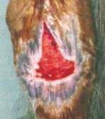
4. Wound six weeks
after skin graft - no
further bandaging
required.
Oliver was left with a
scar but was sound
and ready to return
to work.
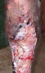
1
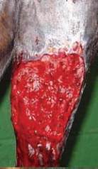
2
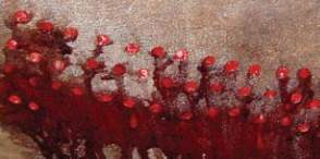
3
4
1. Wound one week after injury.
2. Punch skin grafts were embedded into
granulation tissue.
3. Graft Donor Site with small circles of
skin removed.