Basic HTML Version


WOUND H E A L I NG
3
EQUINE MATTERS
Veterinary Surgeon
Dave Rowlands
XLVets Practice
Penbode Veterinary
Group
F
ailure to manage wounds correctly can result in delayed or poor healing
and increased costs for the owner. In cases where other structures e.g.
joints or tendon sheaths are involved, failure to recognise and treat may
result in euthanasia if infection (sepsis) becomes established.
Wound Healing
Dave Rowlands BVSc CertEM (StudMed) MRCVS,
Penbode Veterinary Group
Equine skin
Skin is the largest organ in the body. In the
horse it varies from 1 - 6mm in thickness. It is
thickest in areas where the chance of injury is
greatest i.e. at the mane and tail attachment,
croup and back. It is thinnest where most
sensitivity is required i.e. the lower and middle
surfaces of the body and limbs. Normal skin
tension is due to elastic fibres in the dermis,
they are the reason why edges retract when
skin is cut.
1. Vascular phase
Wound Healing
- the phases
Initially there is a temporary shutting down
of injured blood vessels (vasoconstriction).
After a few minutes the blood vessels dilate.
A fibrin (protein) seal creates a meshwork
base. Platelets then aggregate and release
hormones and enzymes that cause bleeding
to stop and a clot to form.
2. Inflammatory phase
White blood cells are released into the
damaged tissue and engulf bacteria and
debris. They release growth factors to further
assist wound healing.
4. Granulation and fibroplasia
Granulation tissue consists of invading
blood vessels, fibroblasts and products from
fibroblasts e.g. collagen and elastin. It is
produced 3-4 days after wounding and
serves to rapidly fill in the skin defect, later
the epithelial cells will cover the granulation
tissue to complete healing.
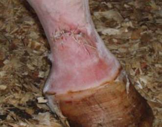
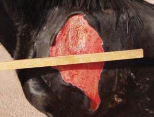
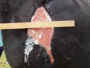
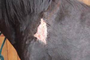
5. Wound contraction
This occurs maximally 5-15 days after
injury. The extent varies according the
wound location. Wounds on the body
with lots of spare skin can undergo large
amounts of contraction as shown in
Figures 1-3 below.
3. Re-epithelialisation
Epithelial cells (superficial skin cells) at the
wound margin migrate across the fibrin
meshwork in the first 24 hours.
A characteristic pink rim of epithelium is
visible after 4-6 days. These cells are very
delicate and migrate slowly, they operate
best in moist conditions free from infection.
Wounds with a narrow gap between the
wound edges can heal fairly quickly by
epithelialisation alone.
Wound healing relies on a complex
series of biochemical reactions. The aim
of wound healing is to restore normal
physical form, structure and function.
Primary Closure
Figure 1
Figure 2
Figure 3